678
Views
9
CrossRef citations to date
0
Altmetric
Original Articles

Impact of heterophil granulocyte depletion caused by 5-fluorouracil on infectious bursal disease virus infection in specific pathogen free chickens

, , , , , & show all
Pages 341-348 | Received 28 Dec 2005, Published online: 18 Jan 2007

Figures & data

Table 1.  Heterophile granulocytes in blood and bursa lesion score

Figure 1. Reactions to IBDV infection in 5-FU-pretreated and non-pretreated chickens. 1a: 5-FU and IBDV inoculation (group 3): 3 days after IBDV inoculation (p.i.), folded surface epithelium was observed (arrows); the medullary epithelial reticular cells form “empty holes”. Anticytokeratin immunostaining. Magnification, 40×. 1b: IBDV inoculation (group 2): 2 days p.i., B-cell depletion. Depletion started in the centre of the medulla and inner part of the cortex. Bu-1b staining. Magnification, 35×. 1c: IBDV inoculation (group 2): 2 days p.i., the follicular cortex flooded by heterophil granulocytes (arrows). Semithin section stained with toluidin blue. Magnification, 500×. 1d: 5-FU and IBDV inoculation (group 3): 2 days p.i., reduction in the number of heterophil granulocytes (arrows). Compare with 1c. Semithin section stained with toluidin blue. Magnification, 500×. 1e: IBDV inoculation (group 2): 2 days p.i., huge numbers of vimentin-positive cells in the medulla and surface epithelium of the follicles. Magnification, 140×. 1f: 5-FU and IBDV inoculation (group 3): 2 days p.i., the number of vimentin-positive cells in the medulla reduced as compared with group 2 (see 1e). Magnification, 140×.
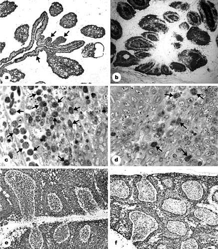
Figure 1.  Reactions to IBDV infection in 5-FU-pretreated and non-pretreated chickens. 1a: 5-FU and IBDV inoculation (group 3): 3 days after IBDV inoculation (p.i.), folded surface epithelium was observed (arrows); the medullary epithelial reticular cells form “empty holes”. Anticytokeratin immunostaining. Magnification, 40×. 1b: IBDV inoculation (group 2): 2 days p.i., B-cell depletion. Depletion started in the centre of the medulla and inner part of the cortex. Bu-1b staining. Magnification, 35×. 1c: IBDV inoculation (group 2): 2 days p.i., the follicular cortex flooded by heterophil granulocytes (arrows). Semithin section stained with toluidin blue. Magnification, 500×. 1d: 5-FU and IBDV inoculation (group 3): 2 days p.i., reduction in the number of heterophil granulocytes (arrows). Compare with 1c. Semithin section stained with toluidin blue. Magnification, 500×. 1e: IBDV inoculation (group 2): 2 days p.i., huge numbers of vimentin-positive cells in the medulla and surface epithelium of the follicles. Magnification, 140×. 1f: 5-FU and IBDV inoculation (group 3): 2 days p.i., the number of vimentin-positive cells in the medulla reduced as compared with group 2 (see 1e). Magnification, 140×.

Table 2.  Histological development in the bursa of Fabricius 5 to 8 days after 5-FU treatment (0 to 3 days after IBDV inoculation)

Figure 2. BSDCs and T cells during IBDV infection. 2a: 5-FU and IBDV inoculation (group 3): 2 days after IBDV infection. The cell contains apoptotic lymphocytes (A) and two demarcated bodies, in which the virus particles are associated with an electron-dense substance (arrow). Part of one of the demarcated bodies is outlined and shown in 2b. Lipid droplets (L) in the cytoplasm. Magnification, 10 000×. 2b: Detail from 2a. In the demarcated body, the virus particles are intermingled with electron dense substance. Magnification, 40 000×. 2c: IBDV inoculation (group 2): 3 days p.i., CD3-positive T cells aggregate in the follicular cortex. Medulla contains only scattered T cells. Magnification, 140×. 2d: IBDV inoculation (group 2): 2 days p.i., the 74.3-positive BSDCs aggregate along the cortico-medullary border in several follicles. Magnification, 160?×. 2e: IBDV inoculation (group 2): 3 days p.i., the 74.3-positive BSDCs seem to be completely aggregated in the medulla. Magnification, 140×. 2f: IBDV inoculation (group 2): 2 days p.i., the anti-IgG staining confirm observations obtained by 74.3 monoclonal antibodies. The IgG-positive BSDCs aggregate along the cortico-medullary border. Compare with 2d. Magnification, 70×.
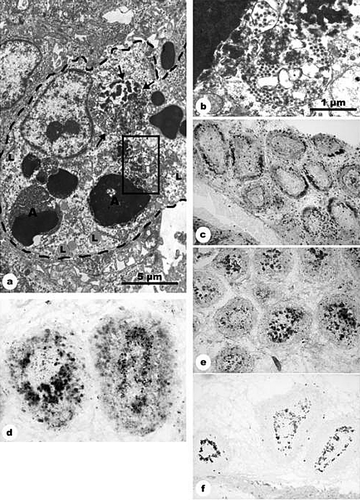
Figure 2.  BSDCs and T cells during IBDV infection. 2a: 5-FU and IBDV inoculation (group 3): 2 days after IBDV infection. The cell contains apoptotic lymphocytes (A) and two demarcated bodies, in which the virus particles are associated with an electron-dense substance (arrow). Part of one of the demarcated bodies is outlined and shown in 2b. Lipid droplets (L) in the cytoplasm. Magnification, 10 000×. 2b: Detail from 2a. In the demarcated body, the virus particles are intermingled with electron dense substance. Magnification, 40 000×. 2c: IBDV inoculation (group 2): 3 days p.i., CD3-positive T cells aggregate in the follicular cortex. Medulla contains only scattered T cells. Magnification, 140×. 2d: IBDV inoculation (group 2): 2 days p.i., the 74.3-positive BSDCs aggregate along the cortico-medullary border in several follicles. Magnification, 160?×. 2e: IBDV inoculation (group 2): 3 days p.i., the 74.3-positive BSDCs seem to be completely aggregated in the medulla. Magnification, 140×. 2f: IBDV inoculation (group 2): 2 days p.i., the anti-IgG staining confirm observations obtained by 74.3 monoclonal antibodies. The IgG-positive BSDCs aggregate along the cortico-medullary border. Compare with 2d. Magnification, 70×.

Reprints and Corporate Permissions

Please note: Selecting permissions does not provide access to the full text of the article, please see our help page How do I view content?

To request a reprint or corporate permissions for this article, please click on the relevant link below:

Academic Permissions

Please note: Selecting permissions does not provide access to the full text of the article, please see our help page How do I view content?

Obtain permissions instantly via Rightslink by clicking on the button below:

If you are unable to obtain permissions via Rightslink, please complete and submit this Permissions form. For more information, please visit our Permissions help page.