Figures & data
Figure 1. Detection of ARV σC-encoding gene in ARV-infected chicken tissues by nested PCR. The σC-encoding gene of ARV in ARV-infected chicken tissues, heart, kidney, bursa, liver, tendon, and intestine (upper and lower panels; lanes 1, 3, 5, 7, 9, and 11) was detected by nested PCR. The expected sizes of first PCR (upper panel) and second PCR (lower panel) products were 330 base pairs and 239 base pairs, respectively. Mock-infected chicken tissues, heart, kidney, bursa, liver, tendon, and intestine used as negative controls (upper and lower panels; lanes 2, 4, 6, 8, 10, and 12). Lane M, Bio 100 DNA ladder™ molecular weight marker.
Figure 2. DNA fragmentation analysis in ARV-infected chicken tissues. Apoptosis induction by ARV S1133 in chicken tissues, heart, kidney, bursa (upper panel; lanes 2, 4, and 6), liver, tendon, and intestine (lower panel; lanes 2, 4, and 6) was detected by DNA fragmentation analysis. Mock-infected chicken tissues, heart, kidney, bursa (upper panel; lanes 1, 3, and 5), liver, tendon, and intestine (lower panel; lanes 1, 3, and 5) were used as negative controls. The chromosomal DNA was separated on a 1.5% agarose gel.
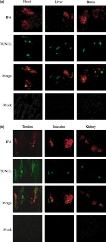
Figure 3. Dual-labelling assay for ARV antigens and apoptosis in ARV-infected tissue sections. Apoptosis induced by ARV in chicken tissues, heart, liver, and bursa (3a) and in tendon, intestine, and kidney (3b) was detected by dual-labelling assay. Extensive syncytium formation was observed in chicken tissues infected with ARV S1133. The dual-labelling (IFA; red) and (TUNEL; green) results (merge) were obtained by SPOT software version 4.5. Mock-infected chicken tissues (mock) were used as negative controls and showed no signals.