2,955
Views
11
CrossRef citations to date
0
Altmetric
ORIGINAL ARTICLES

Infection of HeLa cells by avian infectious bronchitis virus is dependent on cell status

, , , , &
Pages 269-274 | Published online: 09 Jul 2007

Figures & data

Figure 1. Cytopathic effect in IBV-infected HeLa cells. Confluent HeLa monolayers in 100-ml cell culture flasks were inoculated with 1 ml allantoic fluid containing 5×103.4 median egg infectious doses of IBV 41. After 72, CPE was observed. 1a: Mock-infected HeLa cells showing normally growing cells and intact monolayer. 1b: IBV-infected HeLa cells showing CPE: cell rounding, congregating and detaching from the substrate.
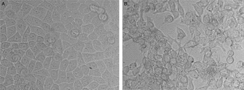
Figure 1.  Cytopathic effect in IBV-infected HeLa cells. Confluent HeLa monolayers in 100-ml cell culture flasks were inoculated with 1 ml allantoic fluid containing 5×103.4 median egg infectious doses of IBV 41. After 72, CPE was observed. 1a: Mock-infected HeLa cells showing normally growing cells and intact monolayer. 1b: IBV-infected HeLa cells showing CPE: cell rounding, congregating and detaching from the substrate.

Figure 2. Growth curve of IBV M41 in HeLa cells. The cell culture supernatant was titrated with ELISA every 6 h post infection until 72 h, when the cells exhibited extensive CPE and detached from the substrate. OD, optical density.
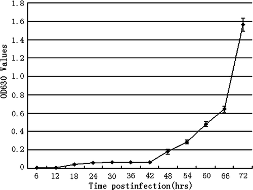
Figure 2.  Growth curve of IBV M41 in HeLa cells. The cell culture supernatant was titrated with ELISA every 6 h post infection until 72 h, when the cells exhibited extensive CPE and detached from the substrate. OD, optical density.

Figure 3. Virus titre (TCID50) change of IBV in HeLa cells with the increasing passage. 3a: IBV M41 was inoculated into freshly dispersed HeLa cells and incubated for 24 passages. The culture supernatant was collected and the TCID50 was tested for every passage. The TCID50 at the level of 103.4±0.2/0.1 ml did not apparently change for the first 12 passages, jumped to 104.2±0.2/0.1 ml for passages 13 to 16, and to 106.6±0.3/0.1 ml for passages 17 to 24 when the experiment was terminated. The experiments were repeated three times. 3b: IBV M41 was inoculated into HeLa cell monolayers and the virus titre averaged 100.9±0.1/0.1 ml over five passages.
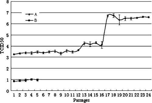
Figure 3.  Virus titre (TCID50) change of IBV in HeLa cells with the increasing passage. 3a: IBV M41 was inoculated into freshly dispersed HeLa cells and incubated for 24 passages. The culture supernatant was collected and the TCID50 was tested for every passage. The TCID50 at the level of 103.4±0.2/0.1 ml did not apparently change for the first 12 passages, jumped to 104.2±0.2/0.1 ml for passages 13 to 16, and to 106.6±0.3/0.1 ml for passages 17 to 24 when the experiment was terminated. The experiments were repeated three times. 3b: IBV M41 was inoculated into HeLa cell monolayers and the virus titre averaged 100.9±0.1/0.1 ml over five passages.

Figure 4. IBV identification by RT-PCR and Western blot assay. 4a: The S1 fragment of the IBV S protein amplified by RT-PCR. The S1 fragment (1025 nucleotides) was amplified from infected allantoic fluid (lane 2) and the fifth passage of IBV in HeLa cells (H5) (lane 3), but not from normal HeLa cells (lane 4) and distilled water (lane 5). Lane 1 represents the DNA ladder (DL2000). 4b: Western blot analysis of IBV propagated in HeLa cells. The viral proteins were separated by sodium dodecyl sulphate-polyacrylamide gel electrophoresis (10%), transferred to nitrocellulose membrane and analysed by western blotting with rabbit antiserum to IBV. Lane 1, purified virus from HeLa cell culture of passage 21; lane 2, passage 8; lane 3, uninfected cell culture. The arrow shows the most abundant viral protein, the nucleoprotein protein (N), with a molecular mass of 45 kDa.
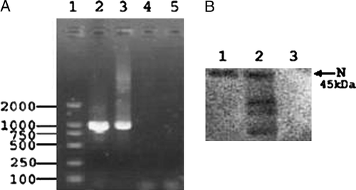
Figure 4.  IBV identification by RT-PCR and Western blot assay. 4a: The S1 fragment of the IBV S protein amplified by RT-PCR. The S1 fragment (1025 nucleotides) was amplified from infected allantoic fluid (lane 2) and the fifth passage of IBV in HeLa cells (H5) (lane 3), but not from normal HeLa cells (lane 4) and distilled water (lane 5). Lane 1 represents the DNA ladder (DL2000). 4b: Western blot analysis of IBV propagated in HeLa cells. The viral proteins were separated by sodium dodecyl sulphate-polyacrylamide gel electrophoresis (10%), transferred to nitrocellulose membrane and analysed by western blotting with rabbit antiserum to IBV. Lane 1, purified virus from HeLa cell culture of passage 21; lane 2, passage 8; lane 3, uninfected cell culture. The arrow shows the most abundant viral protein, the nucleoprotein protein (N), with a molecular mass of 45 kDa.

Reprints and Corporate Permissions

Please note: Selecting permissions does not provide access to the full text of the article, please see our help page How do I view content?

To request a reprint or corporate permissions for this article, please click on the relevant link below:

Academic Permissions

Please note: Selecting permissions does not provide access to the full text of the article, please see our help page How do I view content?

Obtain permissions instantly via Rightslink by clicking on the button below:

If you are unable to obtain permissions via Rightslink, please complete and submit this Permissions form. For more information, please visit our Permissions help page.