Figures & data
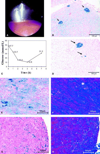
Figure 1. Haemosiderosis (1a to 1d) in a chestnut-fronted macaw (A. severa) and (1e to 1h) in a military macaw (A. militaris) with concurrent diabetes mellitus. 1a: Endoscopic view of the liver from the left caudal thoracic air sac. A miliary distribution of yellow foci is visible on the hepatic surface. 1b: Liver biopsy at presentation showing coarse focal accumulation of iron within macrophages (blue staining, arrows), as well as diffuse iron staining within hepatocytes throughout the parenchyma (Perl's stain). 1c: Blood glucose curve following intramuscular administration of 0.55 IU/kg porcine PZI insulin. The plasma glucose nadir typically occurs between 1 and 3 h after administration, and the effect lasts approximately 8 h. 1d: Necropsy sample of liver collected 24 months after initial presentation. The bird had received eight courses of the iron chelator deferoxamine and its diet had been changed to a low-iron content pellet. Some coarse aggregates are still present (arrows) but iron staining of the hepatocytes is much reduced compared with (1b) (Perl's stain). 1e: Liver biopsy showing marked haemosiderosis (Perl's stain). 1f: A parallel section to (1e) showing increased collagen deposition (blue areas; Masson's Trichrome stain). 1g: Pancreatic biopsy showing moderate haemosiderosis (Perl's stain). 1h: A parallel section to (1g) showing increased in collagen deposition within the pancreatic interstitium (Masson's Trichrome stain).