Figures & data
Figure 1. Ultrasonographic photography of the eye of a 3-week-old affected animal (1b) and its control (1a). In the affected animal, an abnormal hyperechogenicity in the lens cortex and a microphakia are detectable when compared with the control. C, cornea. Le, lens; V, vitreous, Continuous doubleheaded arrow, antero-posterior axis of the lens; discontinuous doubleheaded arrow, antero-posterior axis of the vitreous; arrowhead, with pecten (P); thin arrow, probe-related artefact. M, medial; L, lateral. B-scan/10 MHz.
Figure 2. Section of the eye from a 1-day-old duck: (a) low magnification and (b) high magnification. The anterior cortex and the nucleus of the lens (Le) were replaced by an amorphous eosinophilic material (fibre liquefaction), forming a cavity (*). Lenticular epithelial cells showed cytoplasm vacuolation and ballooning degeneration. C, cornea; Ci, ciliary bodies; Ir, iris; V, vitreous. Haematoxylin–eosin–saffron staining.
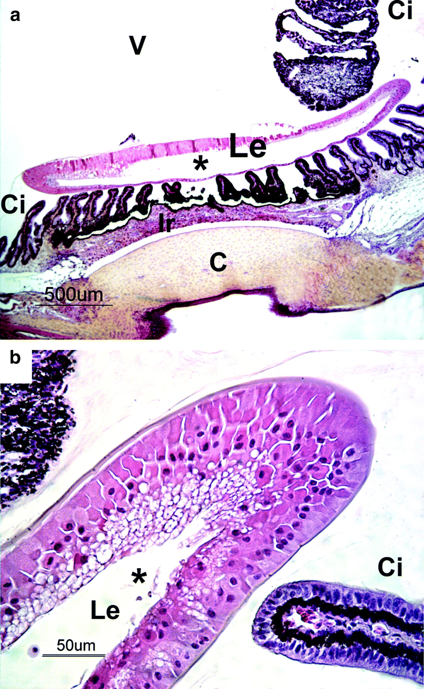
Figure 3. Pre-equatorial semi-thin section of the lens from a 4-week-old duck. Vacuolated cells lined a cavity (*) in the anterior part of the lens containing liquefactive material. Lens fibres were irregular in diameter and dishomogeneous. A degenerated cell (d) is present within the cavity. Toluidine blue staining.