Figures & data
Figure 1. Effect of pretreatment of cells with neuraminidase on the infection by different strains of IBV. Chick embryo kidney cells were incubated in the presence (open boxes) or absence (black boxes) of neuraminidase (NA) of C. perfringens and then infected by one of the four IBV strains Beaudette (Bd), 4/91, Italy 02, and QX at a multiplicity suitable for a plaque assay. The reduction in the plaque number was used to determine the effect of the enzyme treatment on the infectivity of IBV.
Figure 2. Effect of neuraminidase treatment on the infection of TOCs by different strains of IBV. TOCs were incubated in the presence or absence of neuraminidase (NA) from C. perfringens and then infected by one of the four IBV strains (2a) Beaudette (Bd), (2b) 4/91, (2c) QX, and (2d) Italy 02, or mock-infected. Up to 5 days post inoculation, TOCs were analysed for ciliary activity at daily intervals.
Figure 3. Immunofluorescence analysis of cryosections prepared from IBV-infected tracheal organ cultures. At 24 h post infection, sections were stained with an anti-β-tubulin antibody to detect cilia (red) and with a monoclonal anti-N antibody to vizualize virus antigen (green). Bd, Beaudette.
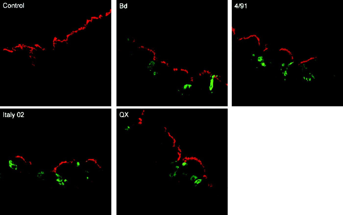
Figure 4. Immunofluorescence analysis of cryosections prepared from IBV-infected tracheal organ cultures. At 24 h post inoculation, sections were stained for goblet cells using an anti-Muc5AC antibody (red) and for virus antigen using a polyclonal IBV serum (green). Areas of co-staining are indicated by white arrows. Bd, Beaudette.
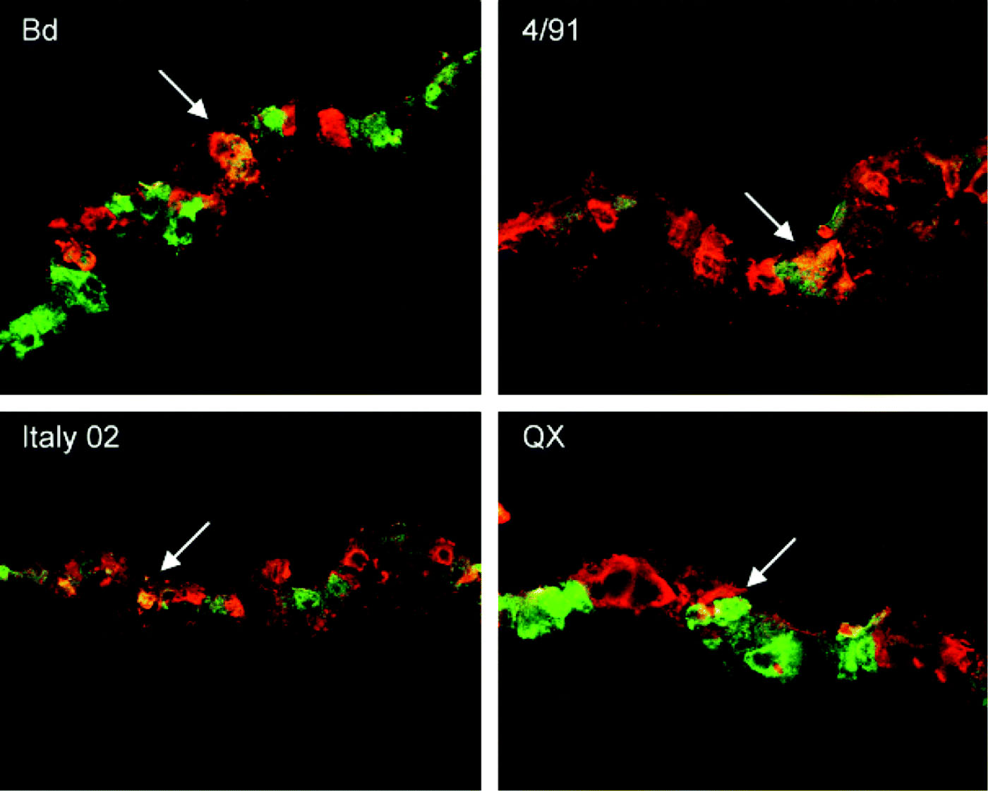
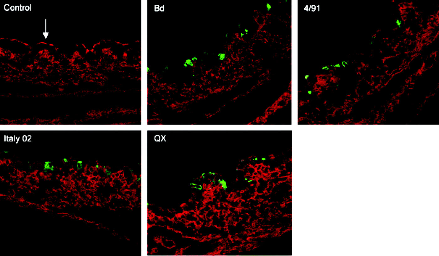
Figure 5. Immunofluorescence analysis of cryosections prepared from IBV-infected tracheal organ cultures. At 24 h post infection, sections were incubated with the lectin MAAII to detect α2,3-linked sialic acid (red). The apical side of the ciliated epithelium is indicated by an arrow. Virus antigen was detected with a monoclonal anti-protein N antibody (green). Interestingly, the apical staining with MAAII was always reduced in infected tissues. Bd, Beaudette.