Figures & data
Figure 1. Western blots showing the reaction of M24 positive serum against ABV N-protein. 1a: Purified, cloned ABV N-protein was separated on sodium dodecyl sulphide-polyacrylamide gel electrophoresis (SDS-PAGE). and blotted onto a plyvinylidene difluoride (PVDF) transfer membrane. It was incubated with positive M24 serum as described in the text. The protein has an apparent molecular weight of approximately 45kDa since it is tagged with a 6 kDa histidine tag. 1b: Western blot of an ABV-infected macaw brain separated on SDS-PAGE and incubated with M24 serum. The single reactive band of approximately 38 kDa corresponds to the predicted position of ABV N-protein.
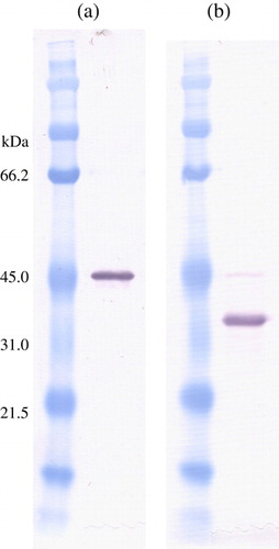
Table 1. Summary of cases, species, tissues available and the results of histopathology and immunohistochemical staining for ABV N-protein
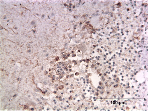
Figure 2. Immunoperoxidase-stained cerebellum of a cockatiel (Case 0427) showing a band of cells staining for ABV N-protein in the Purkinje cell layer. Many of these positive cells are showing extensive cytoplasmic vacuolation. Note that the Purkinje cells themselves do not appear to contain viral protein. Arrows point to stained cells.
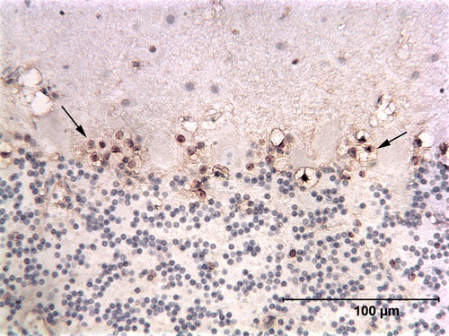
Figure 3. Immunoperoxidase-stained cerebrum of a blue and gold macaw (Case 0437). Cells staining for ABV N-protein are scattered through the cerebral tissue. Note that their nuclei are more heavily stained than their cytoplasm, an expected property of ABV nucleoprotein. Arrows point to stained cells.
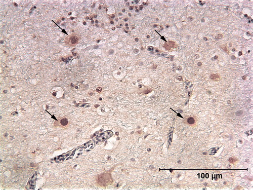
Figure 4. Immunoperoxidase stained spinal cord of a blue and gold macaw (Case 0473) showing the presence of ABV N-protein in neurons and axonal staining within the grey matter.
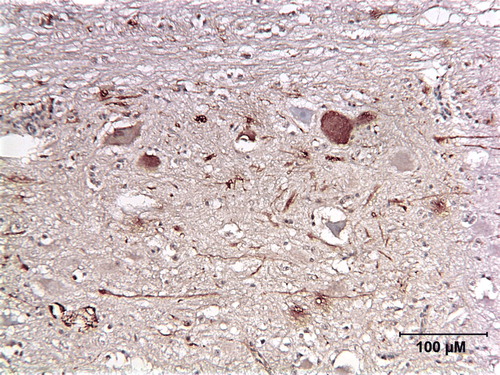
Figure 5. HE-stained cerebrum of a lesser sulphur crested cockatoo (Case 0570). A prominent vascular cuff contains macrophages, plasma cells and lymphocytes.
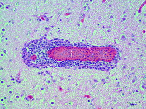
Figure 6. HE-stained cerebellar cortex of a lesser sulphur crested cockatoo (Case 0570). The Purkinje cell layer (large arrows) is devoid of normal Purkinje cells, with the remnant of one degenerated Purkinje cell still present (right arrow). Another cell identified as a neuron has features suggestive of degeneration (small arrow). M, molecular layer; G, granular layer; W, white matter.
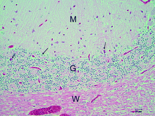
Figure 7. HE-stained cerebellar cortex of an African grey parrot (Case 0387). The Purkinje cell layer of an unaffected (control) animal showing the normal distribution of Purkinje cells (arrows). M, molecular layer; G, granular layer; W, white matter.
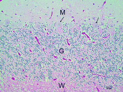
Figure 8. Immunoperoxidase-stained cerebrum of a scarlet macaw (Case 1458) showing multiple cells containing ABV N-protein scattered through the cerebral tissue.
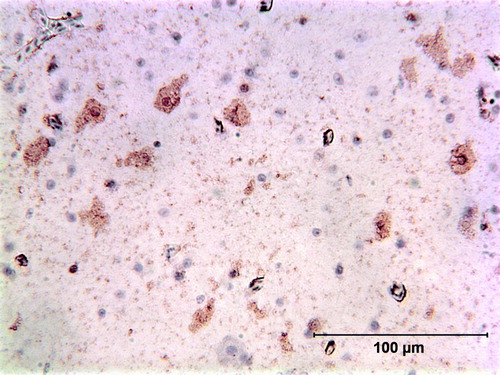
Figure 9. Immunoperoxidase-stained cerebellum of a red-bellied parrot (Case 1583) showing the presence of cells containing ABV N-protein within the Purkinje layer. Purkinje cells appear to be absent or shrunken in this section. Arrows point to stained cells.
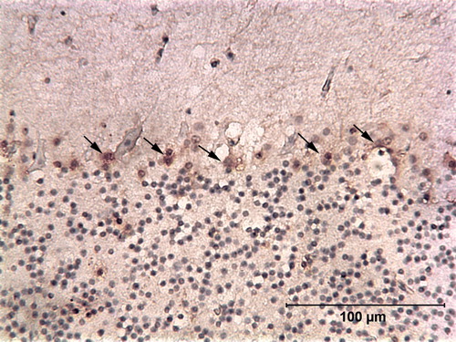
Figure 10. Immunoperoxidase-stained cerebellum of a ruby macaw (Case 1674) showing a thick band of cells containing ABV N-protein associated with the Purkinje cell layer. The Purkinje cells remain unstained. There are also scattered antigen-positive cells throughout the granular layer. Arrows point to stained cells.
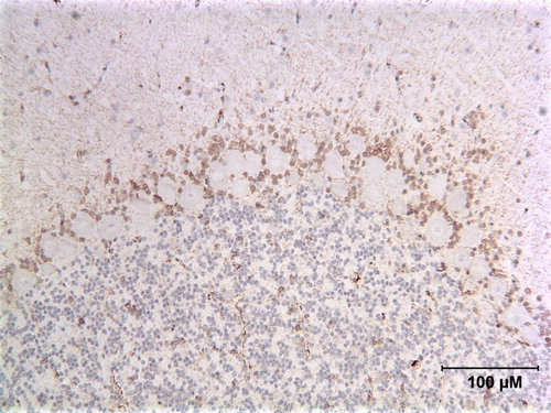
Figure 11. Immunoperoxidase-stained cerebellum of a blue and gold macaw (Case 0382). This bird was not diagnosed originally as suffering from PDD. The band of cells containing ABV N-protein within the Purkinje cell layer, however, is consistent with that seen in confirmed PDD cases. Arrows point to stained cells.