Figures & data
Figure 1. Hungarian partridge with proliferative growths on the upper beaks that surround plastic beak-bits and associated beak necrosis.
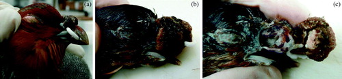
Figure 2. Central circular core of necrotic tissue surrounding the removed plastic bit (empty space), necrotic bone within the beak, and papillary projections of hyperplastic epithelium.
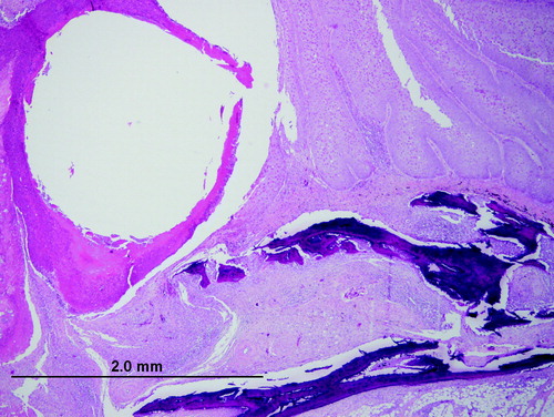
Figure 3. Pox-infected epithelium illustrating hyperplasia, cell swelling, vacuolation, eosinophilic cytoplasmic inclusions and nuclear displacement. Haematoxylin and eosin.
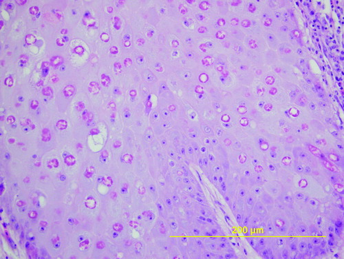
Figure 4. Epithelium from a proliferative mass at the base of the beak surrounded by sheets of poxvirus particles. Electron microscopy magnification 22,000 x at 80 kV.