Figures & data
Table 1. Enzymatic parameters for selected P450 enzymatic activities in duck liver microsomesa.
Figure 1. Effect of selected chemical inhibitors on cytochrome P450-mediated AFB1 epoxide production in duck liver microsomes. Each graph corresponds to six individual birds per experiment (three males and three females). Each graph header indicates the enzymatic activity affected by the inhibitor.
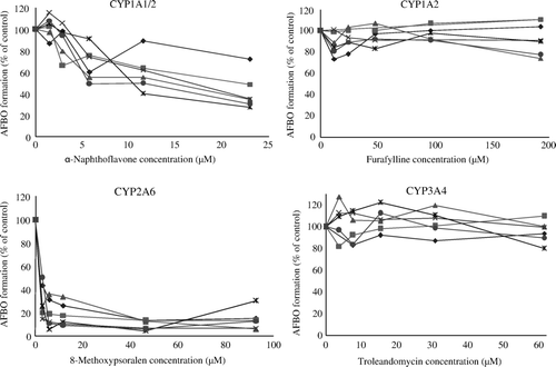
Figure 2. Effect of selected chemical inhibitors on cytochrome P450-mediated AFB1 epoxide production in duck liver microsomes. Each bar corresponds to the mean±standard deviation of six observations per experiment (three males and three females). Each graph header indicates the enzymatic activity affected by the inhibitor. *Significantly different (P < 0.05) from control.
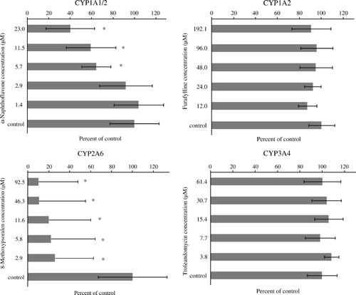
Figure 3. AFB1 epoxidation versus model substrate enzymatic activity in duck liver microsomes (n = 12). The enzyme associated with the model enzymatic activity indicated in the y-axis is shown on top of each graph. A positive significant relationship (P < 0.05) was observed for all enzymatic activities tested.
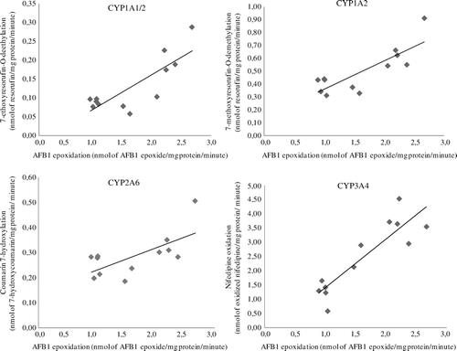
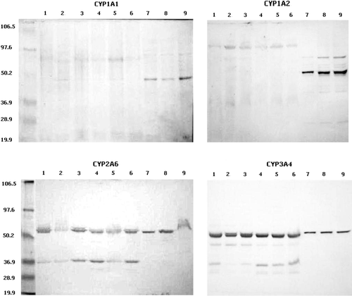
Figure 4. Protein blot of selected duck CYP450 orthologues. Lanes 1 to 6, bands obtained with six different samples of duck microsomes; lanes 7 to 9, microsomes containing cDNA-expressed human CYP450 enzymes as indicated in each graph. Molecular weight standards are shown with their corresponding molecular weight value.