Figures & data
Table 1. C. botulinum strains characterized by PCR and 16S rRNA sequencing.
Figure 1. PFGE patterns of SmaI digests of four avian strains of C. botulinum type C. Lane 1, strain 08-BKT015925; lane 2, 07-BKT002873; lane 3, 07-C6N; and lane 4, 07-V891. The four left lanes in each gel represent samples fixed with formaldehyde, and the four right lines represent samples without. The outermost lanes contain the Lambda Ladder PFG marker (M). 1a: Gel electrophoresed in 0.5x TBE buffer. 1b: Gel electrophoresed in HEPES buffer. The pulse time was ramped from 1 to 40 s for 24 h at (1a) 6 V/cm and (1b) 4 V/cm. Note that the best result appears from fixing cells in formaldehyde and performing electrophoresis in HEPES buffer.
Figure 2. PFGE separation of macrorestriction digests of four avian C. botulinum type C strains using restriction enzymes SmaI, SalI, ApaI and MluI. Lane 1, strain 07-BKT002873; lane 2, 07-V891; lane 3, 07-BKT028387; and lane 4, 08-BKT015925. The outermost lanes contain a low range PFG marker (M). The pulse time was ramped from 1 to 40 s for 30 h at 4 V/cm.
Figure 3. Dendrogram of 15 C. botulinum type C strains based on PFGE digestion patterns obtained by the SalI restriction enzyme. The percentage of similarity of the strains was calculated with the Dice coefficient, and the clustering was performed by the unweighted pair group method using arithmetic averages.
Figure 4. Random-amplified polymorphic-DNA analysis patterns of 15 strains of C. botulinum type C after amplification by primer two (GE Healthcare, Little Chalfont, UK). Lanes 1 to 7, Swedish broiler isolates from 2008; lanes 8 and 10, Swedish broiler isolates from 2007; lane 9, a Norwegian broiler isolate from 2007; lanes 11 to 13, isolates from herring gulls; and lanes 14 and 15, reference strains C-Stockholm and CIP 108943. The outermost lanes are the 100 kb marker (M).
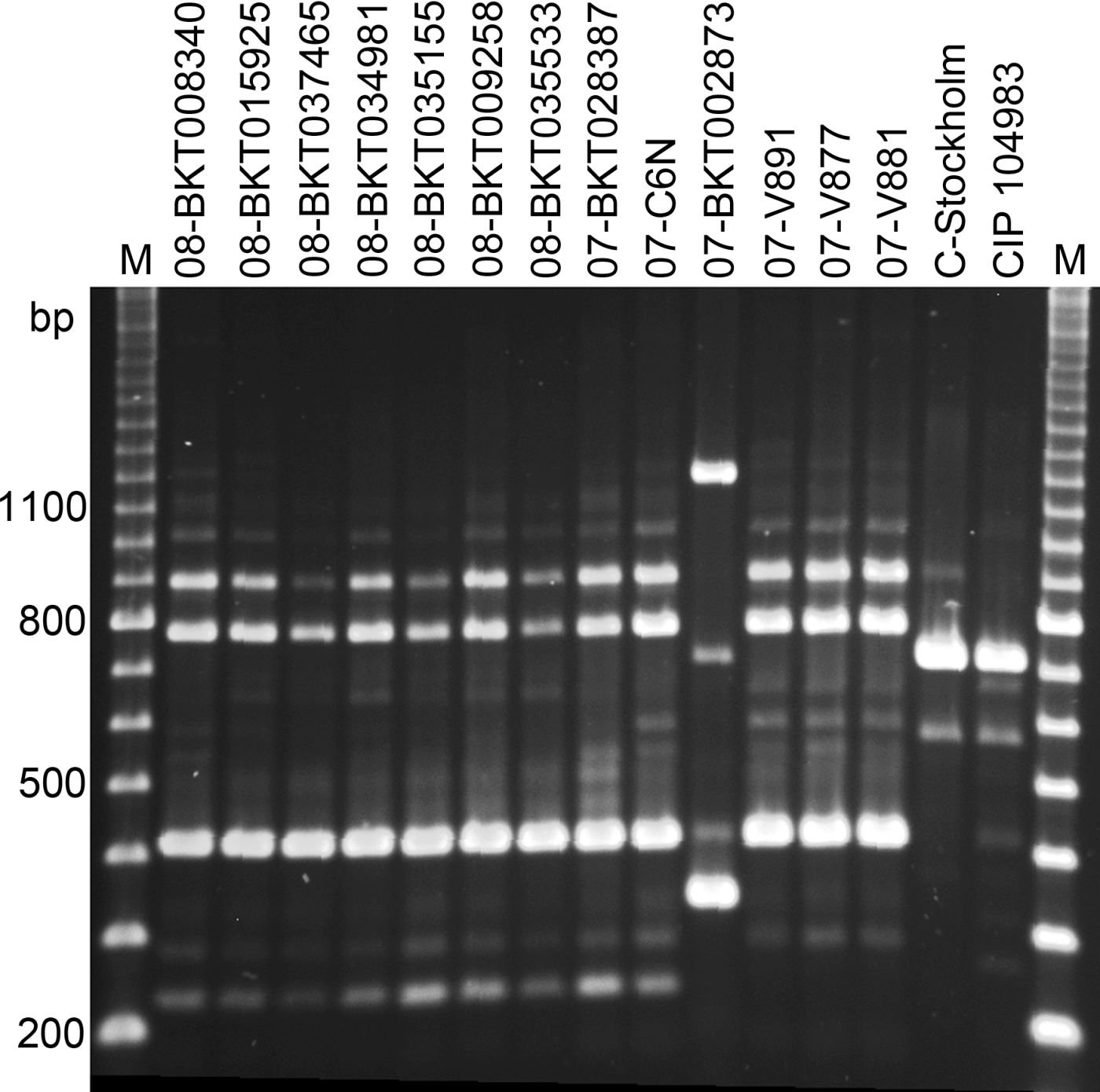
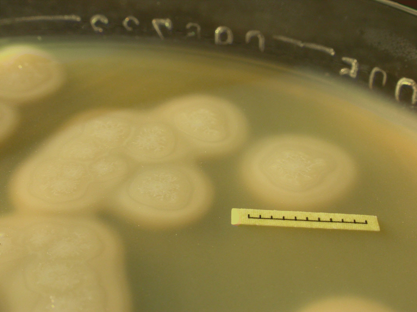
Figure 5. Clostridium botulinum type C, strain 07-BKT028387, in pure culture on McClung Toabe agar.