2,963
Views
32
CrossRef citations to date
0
Altmetric
ORIGINAL ARTICLES

Neurological lesions in chickens experimentally infected with virulent Newcastle disease virus isolates

, , , &
Pages 145-152 | Received 26 Oct 2010, Published online: 15 Apr 2011

Figures & data

Table 1.  Viruses in archived tissue used in the present study.

Figure 1. Schematic diagram showing view of the avian brain according to the actual names. Diagram adapted from Reiner et al. (2004). LSt, lateral striatum; MSt, medial striatum; Nc, nuclei cerebelaris; Mo, medulla oblongata.
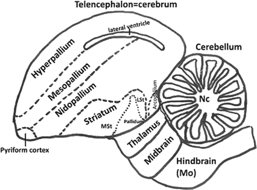
Figure 1.  Schematic diagram showing view of the avian brain according to the actual names. Diagram adapted from Reiner et al. (2004). LSt, lateral striatum; MSt, medial striatum; Nc, nuclei cerebelaris; Mo, medulla oblongata.

Table 2.  Distribution and intensity of histological lesions and stain for virus in different areas of the nervous systema in chickens experimentally infected with different NDV isolates.

Figure 2. Cerebrum (mesopallium), 4-week-old chicken, experimentally infected with Turkey ND strain. 10 d.p.i. Marked infiltration of lymphocytes and plasma cells in and around the vascular wall. Haematoxylin and eosin. Bar=50 µm.
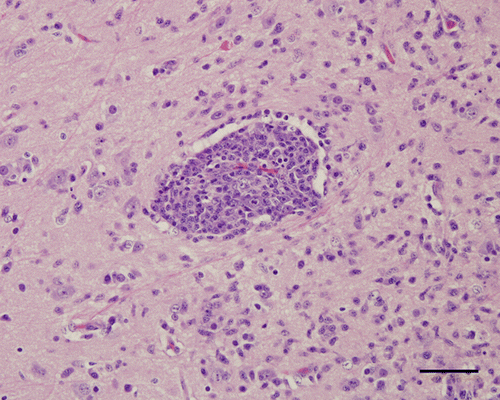
Figure 2.  Cerebrum (mesopallium), 4-week-old chicken, experimentally infected with Turkey ND strain. 10 d.p.i. Marked infiltration of lymphocytes and plasma cells in and around the vascular wall. Haematoxylin and eosin. Bar=50 µm.

Figure 3. Medulla oblongata, 4-week-old chicken, experimentally infected with Nevada cormorant strain. 9 d.p.i. Necrotic neurons undergoing phagocytosis (arrow) by many microglial cells. Haematoxylin and eosin. Bar=50 µm.
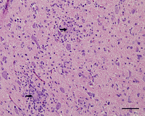
Figure 3.  Medulla oblongata, 4-week-old chicken, experimentally infected with Nevada cormorant strain. 9 d.p.i. Necrotic neurons undergoing phagocytosis (arrow) by many microglial cells. Haematoxylin and eosin. Bar=50 µm.

Figure 4. Cerebellum, 4-week-old chicken, experimentally infected with Turkey ND strain. 10 d.p.i. The molecular layer (M) has focal encephalitis and vascular proliferation. Some Purkinje cells in the subjacent Purkinje cell layer (P) are missing. In the granular layer (G), there are no changes. Haematoxylin and eosin. Bar=50 µm.
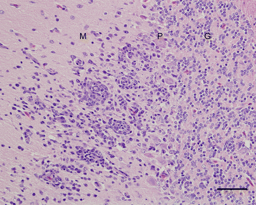
Figure 4.  Cerebellum, 4-week-old chicken, experimentally infected with Turkey ND strain. 10 d.p.i. The molecular layer (M) has focal encephalitis and vascular proliferation. Some Purkinje cells in the subjacent Purkinje cell layer (P) are missing. In the granular layer (G), there are no changes. Haematoxylin and eosin. Bar=50 µm.

Figure 5. Medulla oblongata, 4-week-old chicken, experimentally infected with Turkey ND strain. Spongy changes (*), axonal spheroids (arrow head) and hypertrophy of the nuclei of astrocytes. Haematoxylin and eosin. Bar=50 µm.
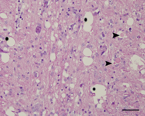
Figure 5.  Medulla oblongata, 4-week-old chicken, experimentally infected with Turkey ND strain. Spongy changes (*), axonal spheroids (arrow head) and hypertrophy of the nuclei of astrocytes. Haematoxylin and eosin. Bar=50 µm.

Figure 6. Hyperpallium, 4-week-old chicken, experimentally infected with Korea 97-147 strain. 5 d.p.i. Immunohistochemistry for viral nucleoprotein (NDV). Multiple granules are identifiable in the cytoplasm of neurons. Bar=50 µm.
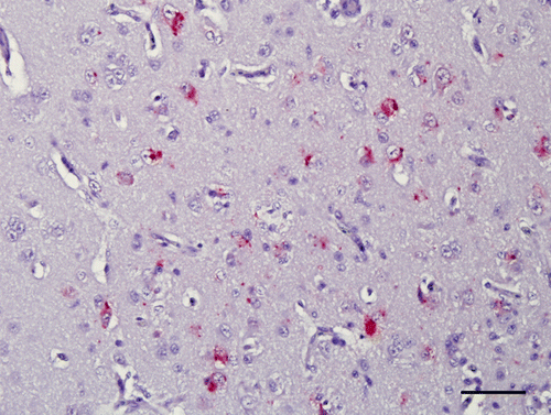
Figure 6.  Hyperpallium, 4-week-old chicken, experimentally infected with Korea 97-147 strain. 5 d.p.i. Immunohistochemistry for viral nucleoprotein (NDV). Multiple granules are identifiable in the cytoplasm of neurons. Bar=50 µm.

Figure 7. Cerebrum (hyperpallium), 4-week-old chicken, experimentally infected with Turkey ND strain. 10 d.p.i. Immunohistochemistry for GFAP. Many GFAP-positive astrocytes can be observed in the areas of inflammation. Bar=50 µm.
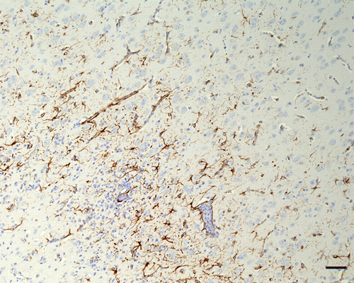
Figure 7.  Cerebrum (hyperpallium), 4-week-old chicken, experimentally infected with Turkey ND strain. 10 d.p.i. Immunohistochemistry for GFAP. Many GFAP-positive astrocytes can be observed in the areas of inflammation. Bar=50 µm.

Figure 8. Spinal cord, 4-week-old chicken, experimentally infected with Turkey ND strain. 10 d.p.i. Immunohistochemistry for GFAP. Grey matter and a focal area under meninges with inflammatory changes and a moderate increase of GFAP expression around. Bar=50 µm.
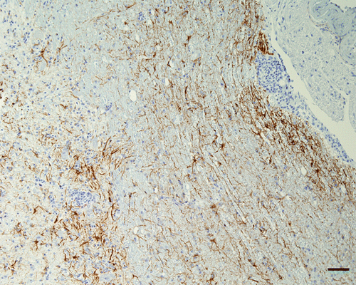
Figure 8.  Spinal cord, 4-week-old chicken, experimentally infected with Turkey ND strain. 10 d.p.i. Immunohistochemistry for GFAP. Grey matter and a focal area under meninges with inflammatory changes and a moderate increase of GFAP expression around. Bar=50 µm.

Table 3.  Greatest histologic severity lesions, degree of inflammation, signal for CD3 and intensity of signal for GFAP.

Reprints and Corporate Permissions

Please note: Selecting permissions does not provide access to the full text of the article, please see our help page How do I view content?

To request a reprint or corporate permissions for this article, please click on the relevant link below:

Academic Permissions

Please note: Selecting permissions does not provide access to the full text of the article, please see our help page How do I view content?

Obtain permissions instantly via Rightslink by clicking on the button below:

If you are unable to obtain permissions via Rightslink, please complete and submit this Permissions form. For more information, please visit our Permissions help page.