Figures & data
Figure 1. Neighbour-joining phylogenetic tree based on the nucleotide sequences of the ORF 1b encoding the polymerase gene of six ANV-1 strains. The numbers near the branches indicate confidence level calculated by bootstrap (n=1000).
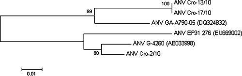
Figure 2. Focal mononuclear cell hyperplasia (lymphocytes) in the kidney. H & E stain (scale bar= 10 µm).
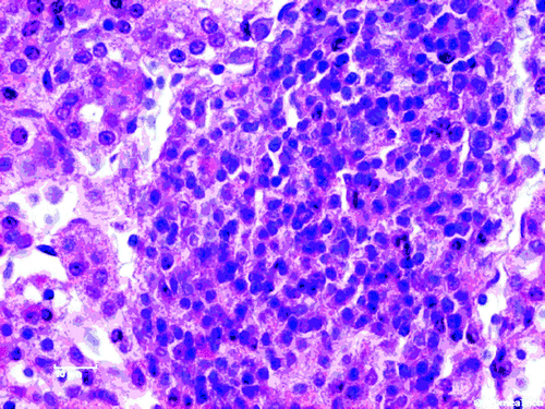
Figure 3. Focal mononuclear cell hyperplasia (lymphocytes and plasma cells) in the kidney. H & E stain (scale bar=10 µm).
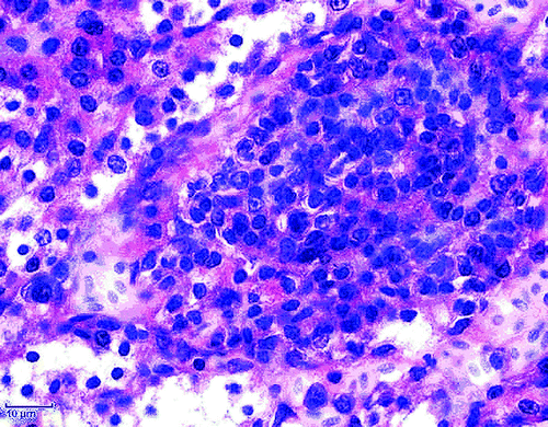
Figure 4. Focal mononuclear cell hyperplasia (plasma cells) in the kidney. H & E stain (scale bar= 10 µm).
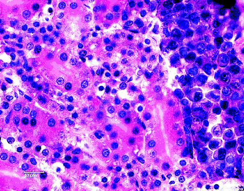
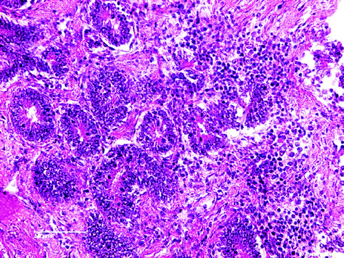
Figure 5. Diffuse pericryptal mononuclear cell hyperplasia (lymphocytes) and fibroplasia in the duodenum. H & E stain (scale bar= 50 µm).