2,504
Views
25
CrossRef citations to date
0
Altmetric
ORIGINAL ARTICLES

Mycoplasma gallisepticum experimental infection and tissue distribution in chickens, sparrows and pigeons

Pages 349-354 | Received 03 Feb 2011, Published online: 04 Aug 2011

Figures & data

Table 1.  HI results for chickens, pigeons and sparrows 21 d.p.i. with M. gallisepticum.

Table 2.  Number of birds positive for M. gallisepticum by culture of 10 birds tested at 3, 7, 14, and 21 d.p.i.

Figure 1. Photomicrographs of IHC staining for M. gallisepticum antigens in tissues from experimentally infected chickens, sparrows, and pigeons. Brown stain indicates presence of M. gallisepticum antigens. Tissues are counterstained using haematoxylin (blue). 1a: Moderate staining (++ ) in a chicken's conjunctiva. 1b: Moderate staining (++) in a chicken's nasal cavity. 1c: Intense staining (+++) in a chicken's trachea. 1d: Moderate staining (++) in a chicken's air sac. 1e: Moderate staining (++) in a sparrow's conjunctiva. 1f: Mild staining (+) in a sparrow's trachea. 1g: Mild staining (+) in a pigeon's conjunctiva. All scale bars = 50 µm.
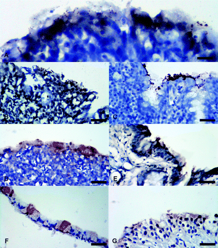
Figure 1.  Photomicrographs of IHC staining for M. gallisepticum antigens in tissues from experimentally infected chickens, sparrows, and pigeons. Brown stain indicates presence of M. gallisepticum antigens. Tissues are counterstained using haematoxylin (blue). 1a: Moderate staining (++ ) in a chicken's conjunctiva. 1b: Moderate staining (++) in a chicken's nasal cavity. 1c: Intense staining (+++) in a chicken's trachea. 1d: Moderate staining (++) in a chicken's air sac. 1e: Moderate staining (++) in a sparrow's conjunctiva. 1f: Mild staining (+) in a sparrow's trachea. 1g: Mild staining (+) in a pigeon's conjunctiva. All scale bars = 50 µm.

Table 3.  Tissues from chickens, sparrows, and pigeons with positive staining for M. gallisepticum antigens using IHC and stain intensity.

Figure 2. Electrophoresis analysis (1.5% agarose gel) of PCR products of M. gallisepticum shedding amplified by Mycoplasma universal primers (Lauerman et al., 1995). Lanes 1 and 9, 100 base pair (bp) DNA marker (Promega Corp, Madison, Wisconsin, USA); lanes 2 to 6, unidentified Mycoplasma sp. (band size around 500 bp) from inoculated pigeon cultures; lane 7, positive M. gallisepticum sample (band size around 900 bp) from inoculated chicken culture; lane 8 = negative control.
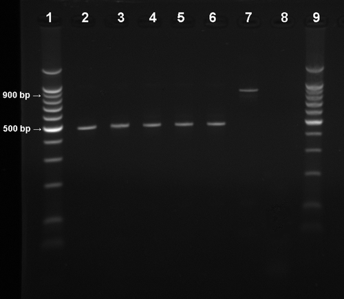
Figure 2.  Electrophoresis analysis (1.5% agarose gel) of PCR products of M. gallisepticum shedding amplified by Mycoplasma universal primers (Lauerman et al., 1995). Lanes 1 and 9, 100 base pair (bp) DNA marker (Promega Corp, Madison, Wisconsin, USA); lanes 2 to 6, unidentified Mycoplasma sp. (band size around 500 bp) from inoculated pigeon cultures; lane 7, positive M. gallisepticum sample (band size around 900 bp) from inoculated chicken culture; lane 8 = negative control.

Reprints and Corporate Permissions

Please note: Selecting permissions does not provide access to the full text of the article, please see our help page How do I view content?

To request a reprint or corporate permissions for this article, please click on the relevant link below:

Academic Permissions

Please note: Selecting permissions does not provide access to the full text of the article, please see our help page How do I view content?

Obtain permissions instantly via Rightslink by clicking on the button below:

If you are unable to obtain permissions via Rightslink, please complete and submit this Permissions form. For more information, please visit our Permissions help page.