1,241
Views
2
CrossRef citations to date
0
Altmetric
ORIGINAL ARTICLES

Bilateral coxofemoral degenerative joint disease in a juvenile male yellow-eyed penguin (Megadyptes antipodes)

&
Pages 371-375 | Received 11 Jan 2011, Published online: 04 Aug 2011

Figures & data

Figure 1. Post-mortem ventrodorsal radiographs of a juvenile YEP with bilateral coxofemoral degenerative joint disease (top) and unaffected age-matched control (bottom).
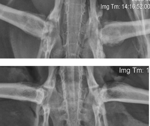
Figure 1.  Post-mortem ventrodorsal radiographs of a juvenile YEP with bilateral coxofemoral degenerative joint disease (top) and unaffected age-matched control (bottom).

Figure 2. Gross formalin-fixed transverse section of the left coxofemoral joint of a juvenile YEP with bilateral coxofemoral degenerative joint disease. Note the spinal cord at the top (arrow), and the femoral shaft extending down and to the left. The femoral head remnant is surrounded by necrotic debris, collagen, and organizing haemorrhage. The horizontal crack in the section is artefactual. Scale bar = 1 cm.
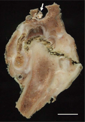
Figure 2.  Gross formalin-fixed transverse section of the left coxofemoral joint of a juvenile YEP with bilateral coxofemoral degenerative joint disease. Note the spinal cord at the top (arrow), and the femoral shaft extending down and to the left. The femoral head remnant is surrounded by necrotic debris, collagen, and organizing haemorrhage. The horizontal crack in the section is artefactual. Scale bar = 1 cm.

Figure 3. The defleshed right proximal femur of a juvenile YEP with bilateral coxofemoral degenerative joint disease (left) and an unaffected age-matched control (right). The affected femoral head is markedly shrunken, angular, and roughened, with osteophyte formation on the femoral neck, and greater and lesser trochanters. Scale bar = 1 cm.
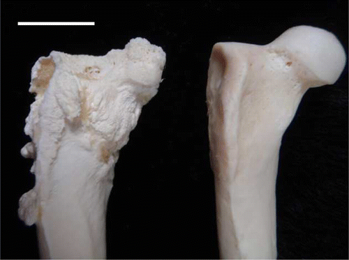
Figure 3.  The defleshed right proximal femur of a juvenile YEP with bilateral coxofemoral degenerative joint disease (left) and an unaffected age-matched control (right). The affected femoral head is markedly shrunken, angular, and roughened, with osteophyte formation on the femoral neck, and greater and lesser trochanters. Scale bar = 1 cm.

Figure 4. The defleshed right pelvis of a juvenile YEP with bilateral coxofemoral degenerative joint disease (top) with an unaffected age-matched control (bottom). The acetabulum is markedly widened and there is circumferential osteophyte formation.
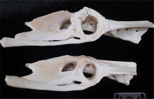
Figure 4.  The defleshed right pelvis of a juvenile YEP with bilateral coxofemoral degenerative joint disease (top) with an unaffected age-matched control (bottom). The acetabulum is markedly widened and there is circumferential osteophyte formation.

Figure 5. Histology of the left femoral head of a juvenile YEP with bilateral coxofemoral degenerative joint disease (haematoxylin and eosin), with the remains of the proximal femoral head at the top left (F). The articular cartilage is absent, and much of the normal epiphyseal bone has been resorbed. The remaining trabecular bone is covered by a thick layer of mature lamellar collagen (C). Fibrin and cellular debris occupy pockets within the remaining joint space (arrow). Note the florid periosteal reaction that forms delicate trabeculae which protrude nearly perpendicularly from the periostium (P). Scale bar = 1000 µm.
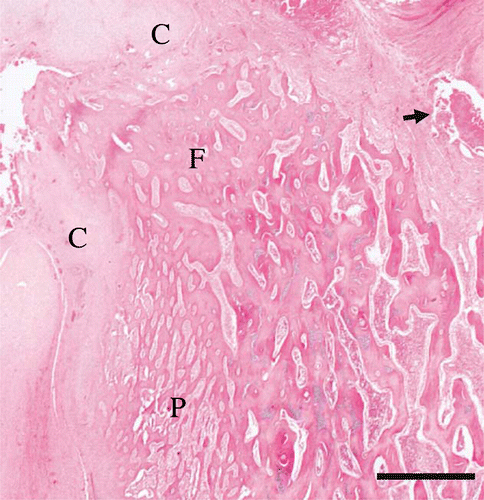
Figure 5.  Histology of the left femoral head of a juvenile YEP with bilateral coxofemoral degenerative joint disease (haematoxylin and eosin), with the remains of the proximal femoral head at the top left (F). The articular cartilage is absent, and much of the normal epiphyseal bone has been resorbed. The remaining trabecular bone is covered by a thick layer of mature lamellar collagen (C). Fibrin and cellular debris occupy pockets within the remaining joint space (arrow). Note the florid periosteal reaction that forms delicate trabeculae which protrude nearly perpendicularly from the periostium (P). Scale bar = 1000 µm.

Figure 6. Histology of the articular surface of the left femoral head of a juvenile YEP with bilateral coxofemoral degenerative joint disease (haematoxylin and eosin). The cortical bone and articular cartilage are absent, and remaining superficial trabecular bone has been covered by maturing sheets of collagen interspersed with fragments of bone (arrows), with layers of superficial fibrin (F). Within intact trabecular bone there is active resorption by many osteoclasts, and Howship's lacunae are prominent (arrowheads). Scale bar = 150 µm.
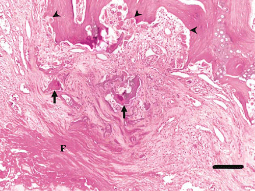
Figure 6.  Histology of the articular surface of the left femoral head of a juvenile YEP with bilateral coxofemoral degenerative joint disease (haematoxylin and eosin). The cortical bone and articular cartilage are absent, and remaining superficial trabecular bone has been covered by maturing sheets of collagen interspersed with fragments of bone (arrows), with layers of superficial fibrin (F). Within intact trabecular bone there is active resorption by many osteoclasts, and Howship's lacunae are prominent (arrowheads). Scale bar = 150 µm.

Reprints and Corporate Permissions

Please note: Selecting permissions does not provide access to the full text of the article, please see our help page How do I view content?

To request a reprint or corporate permissions for this article, please click on the relevant link below:

Academic Permissions

Please note: Selecting permissions does not provide access to the full text of the article, please see our help page How do I view content?

Obtain permissions instantly via Rightslink by clicking on the button below:

If you are unable to obtain permissions via Rightslink, please complete and submit this Permissions form. For more information, please visit our Permissions help page.