Figures & data
Figure 1. 1A: Mature plexiform-like CVL in the lung of a 24-week-old-broiler from a line selected for susceptibility to IPAH. The CVL occupies the position of an inter-parabronchial arteriole supplying two parabronchi on the ventral surface of the lung (100×). 1B: Foam-type macrophages (arrow) are present around the periphery of a matrix of intimal proliferating cells (arrowhead) at the centre of the CVL (400×).
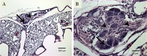
Table 1. Primary antibodies used for immunohistochemistry.
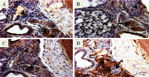
Figure 2. Photomicrographs of immunostained lung sections from IPAH-susceptible broilers showing positive staining for angioproliferative molecules that include (2A) vWF, (2B) αSMA, (2C) VEGF, (2D) VEGFR-2, (2E) HIF-1α, (2F) survivin, (2G) tenascin, (2H) fibronectin, (2I) collagen type III, and (2J) collagen type IV. It must be noted that we examined the lung sections from 8-week-old to 24-week-old broilers. The lung sections presented here are from 12-week-old broilers except for VEGFR-2 (20 weeks old). The brown-coloured reaction product indicates positive staining. For each molecule, the foam-type macrophages are indicated by an arrow and the matrix of intimal proliferating cells by an arrowhead. Original magnification ×400, scale bar =50 µm.
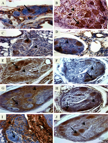
Figure 3. Photomicrographs of immunostained lung sections from IPAH-susceptible broiler chickens showing positive staining for immune/inflammatory cell markers that include (3A) monocytes/macrophages (KUL01), (3B) cytotoxic lymphocytes (CD8), (3C) B cells (Bu-1), and (3D) MHC class II cells (Ia). It must be noted that we examined the lung sections from 8-week-old to 24-week-old broilers. The lung sections presented here are from 12-week-old broilers. The brown-coloured reaction product indicates positive staining. For each immune/inflammatory cell, the foam-type macrophages are indicated by an arrow and matrix of intimal proliferating cells by an arrowhead. Original magnification ×400, scale bar = 50 µm.