Figures & data
Figure 1. Lateral view of a cliff swallow head. There is mild swelling of the eyelids and nictitans, with loss of feathers from the skin of the upper and, to a lesser extent, the lower eyelid and from the skin at the medial canthus.
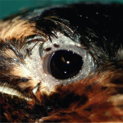
Figure 2. Conjunctiva from a cliff swallow with conjunctivitis. Lymphocytes and plasma cells (inset: bar = 10 µm), along with a focus of follicular lymphoid hyperplasia (*), infiltrate and expand the lamina propria. Bar = 20 µm. Haematoxylin and eosin.
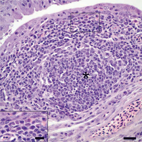
Figure 3. Fornix of the conjunctiva from a cliff swallow with conjunctivitis. There is hyperplasia of conjunctival epithelium, evidenced by tufts and pilings of epithelial cells, and heterophils in low numbers are scattered in the hyperplastic epithelium. Note that the lamina propria is infiltrated by lymphocytes and plasma cells, and few cryptosporidial stages are present along the surfaces of epithelial cells. Bar = 20 µm. Haematoxylin and eosin.
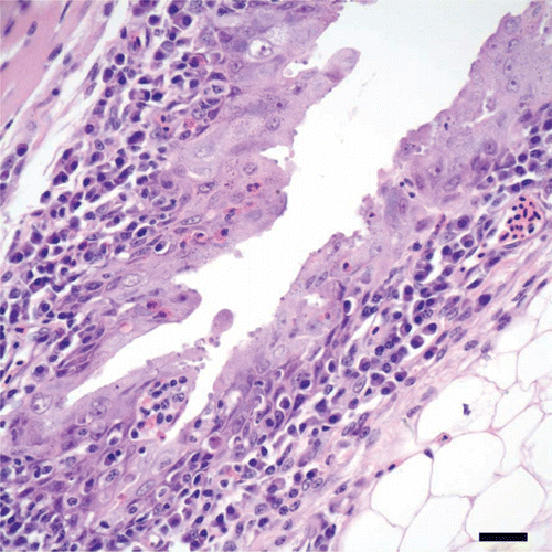
Figure 4. Nasal concha of a cliff swallow with rhinitis. The lamina propria of the nasal mucosa overlying the turbinate (t) is expanded by a lymphoplasmacytic infiltrate with follicular lymphoid hyperplasia. Note the cluster of cyrptosporidial stages along the surface of the epithelium. Bar = 20 µm. Haematoxylin and eosin.
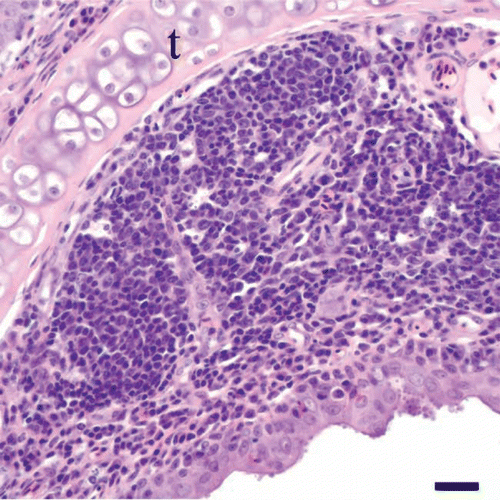
Figure 5. Nasal septum of a cliff swallow with rhinitis. There is hyperplasia of the nasal mucosal epithelium along the nasal septum (s), as evidenced by piling and layering of epithelial cells, together with scattered intra-epithelial heterophils. Note the infiltrate of lymphocytes and plasma cells in the lamina propria and the cryptosporidial stages along the surfaces of epithelial cells. Bar = 20 µm. Haematoxylin and eosin.
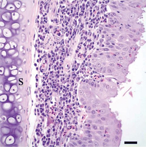
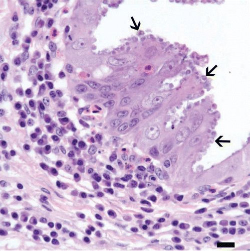
Figure 6. Conjunctiva from a cliff swallow with conjunctivitis. Numerous, round protozoa 2 to 5 µm in diameter, consistent with cryptosporidial developmental stages (arrows), are located in or at the apical margins of epithelial cells along this segment of hyperplastic conjunctival epithelium. An infiltrate of plasma cells with occasional heterophils is present in the underlying connective tissue of the lamina propria. Bar = 10 µm. Haematoxylin and eosin.