Figures & data
Table 1. Details of all cases of extra-intestinal coccidiosis in kiwi (Apteryx spp.) diagnosed at Massey University (1991 to 2011) using histological methods.
Table 2. Clinical signs reported in kiwi (Apteryx spp.) associated with various forms of extra-intestinal coccidiosis.
Figure 1. 1a: Partially eviscerated juvenile brown kiwi (A. mantelli; Case 1976) with kidneys in situ demonstrating renal pallor and renomegally associated with a heavy burden of coccidia. BW, body wall; CaL, caudal pole left kidney; CaR, caudal pole right kidney; CrL, cranial pole left kidney; CrR, cranial pole right kidney; R, transected rectum. 1b: Low-power view illustrating coccidial organisms (arrow) within collecting ducts of the medullary cone in the kidney of a juvenile brown kiwi (H&E stain). Scale bar = 200 µm. 1c: Coccidial oocysts causing dilation and obstruction of a collecting duct in the kidney of a juvenile brown kiwi (H&E stain). Scale bar = 20 µm. 1d: Renal collecting ducts of a juvenile brown kiwi demonstrating gametocytes within epithelial cells (H&E stain). Scale bar = 20 µm. 1e: Immature meronts causing tubular dilation within collecting duct epithelial cells in the kidney of a juvenile brown kiwi (H&E stain). Scale bar = 20 µm. 1f: Mature meronts with radially projecting merozoites within a renal collecting duct of a juvenile brown kiwi (H&E stain). Scale bar = 20 µm.
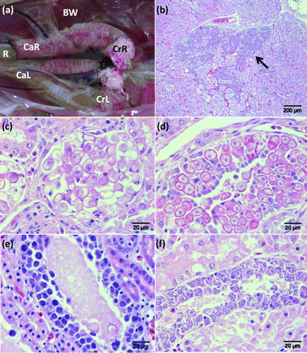
Figure 2. 2a: Pinpoint white serosal foci associated with coccidiosis in the liver of a brown kiwi chick (Case 2586). Scale bar = 20 mm. 2b: Focal areas of necrosis within the liver of a juvenile brown kiwi associated with coccidial meronts (H&E stain). Scale bar = 500 µm. 2c: An immature macromeront within the liver of a juvenile brown kiwi (H&E stain). Scale bar = 200 µm. 2d: An alternative form of macromeront surrounded by a thick eosinophilic capsule within the liver of a brown kiwi chick (H&E stain). Scale bar = 200 µm.
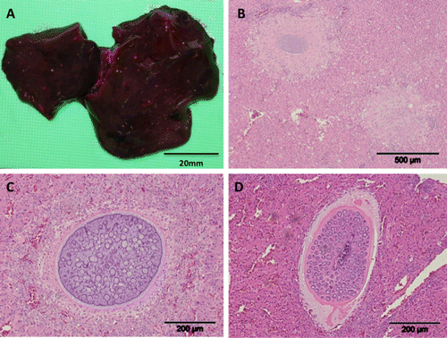
Table 3. Morphometrics of various extra-intestinal coccidial stages identified in this study.
Figure 3. A macromeront within the spleen of a juvenile brown kiwi (H&E stain). Scale bar = 100 µm.
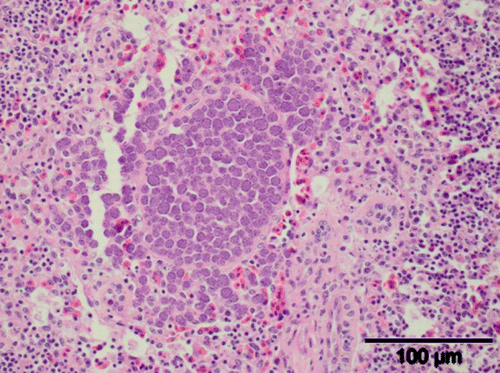
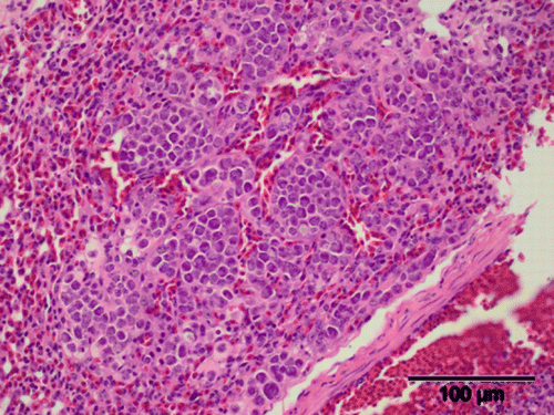
Figure 4. Pulmonary histopathology of a juvenile brown kiwi illustrating coccidial meronts (H&E stain). Scale bar = 100 µm.