1,681
Views
3
CrossRef citations to date
0
Altmetric
ORIGINAL ARTICLES

A case of cutaneous angiolipoleiomyoma (angiomyolipoma) in a budgerigar (Melopsittacus undulatus)

, , , &
Pages 511-515 | Received 19 Mar 2013, Accepted 24 Jun 2013, Published online: 10 Oct 2013

Figures & data

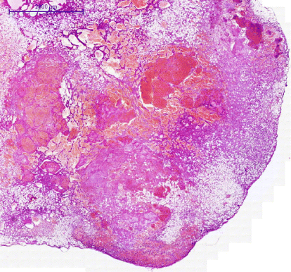
Figure 1. Histopathological picture of the budgerigar angiolipoleiomyoma with classical triads—haemangioma, smooth muscle bundles, and mature white adipose tissues—at low magnification. Haematoxylin and eosin. Bar: 2000 µm.
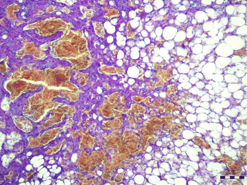
Figure 2. Smooth muscle bundles arranged in well-defined fascicles and around vascular channels. Haematoxylin and eosin. Bar: 100 µm.
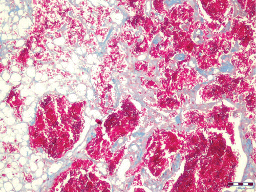
Figure 3. Masson's trichrome stained the smooth muscle portions of the mixed mesenchymal tumour bright red, and the collagen blue. Masson's trichrome. Bar: 100 µm.
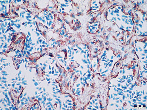
Figure 4. α-SMA cytoplasmic positivity (brown) in benign neoplastic smooth muscle cells of the angiolipoleiomyoma. Immunohistochemistry, diamino-benzidine–chromogen. Bar: 20 µm.
Figure 5. Sarcomeric actin moderate cytoplasmic positivity (brown) in benign neoplastic smooth muscle cells of the angiolipoleiomyoma. Immunohistochemistry, diamino-benzidine–chromogen. Bar: 20 µm.
Figure 6. Myosin weak cytoplasmic positivity (brown) in neoplastic smooth muscle cells of the angiolipoleiomyoma. Immunohistochemistry, diamino-benzidine–chromogen. Bar: 20 µm.
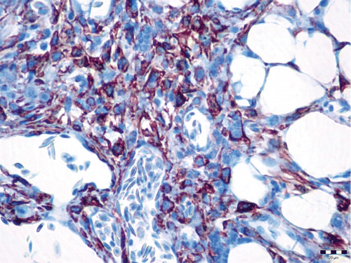
Figure 7. Claudin-5 membranous positivity (brown) in neoplastic endothelial cells of the angiolipoleiomyoma. Immunohistochemistry, diamino-benzidine–chromogen. Bar: 20 µm.

Reprints and Corporate Permissions

Please note: Selecting permissions does not provide access to the full text of the article, please see our help page How do I view content?

To request a reprint or corporate permissions for this article, please click on the relevant link below:

Academic Permissions

Please note: Selecting permissions does not provide access to the full text of the article, please see our help page How do I view content?

Obtain permissions instantly via Rightslink by clicking on the button below:

If you are unable to obtain permissions via Rightslink, please complete and submit this Permissions form. For more information, please visit our Permissions help page.