1,668
Views
17
CrossRef citations to date
0
Altmetric
ORIGINAL ARTICLES

Evaluation of a novel strain of infectious bronchitis virus emerged as a result of spike gene recombination between two highly diverged parent strains

, , , , &
Pages 249-257 | Received 10 Jan 2014, Accepted 23 Mar 2014, Published online: 12 May 2014

Figures & data

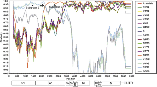
Figure 1. SimPlot analysis of the structural protein gene region of several Australian IBV strains. Structural protein gene order is shown below the graph and is approximately to scale (given different strains can have different gene lengths). The gap between genes M and 5a represents a region with unknown function. Analysis was performed using the SimPlot software (http://sray.med.som.jhmi.edu/SCRoftware/simplot/). Parameters used were a 200 bp window with a 20 bp step, with N1/08 used as the query strain, N1/88 and N1/03 as the reference strains and Armidale as the required “other” strain. The N1/08 3′ crossover point is marked by an arrow, while the representative subgroup 2 and 3 strains are marked by arrowheads.
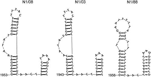
Figure 3. Double stem-loop structure found in the region of the predicted 3′ crossover site. Predictions were made using RNAstructure version 5.1 (http://rna.urmc.rochester.edu/index.html) using the default parameters. The number indicates the location of the first base of the stem loop in relation to the initiation codon of the S gene.
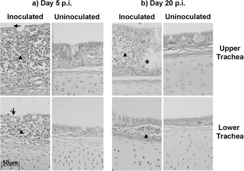
Figure 4. Microscopic lesions found in the tracheas of experimental chickens. 4a: Tracheal sections from chickens at day 5 p.i. 5b: Tracheal sections from chickens at day 20 p.i. The left panels in (a) and (b) show tracheal sections from chickens in the N1/08 inoculated groups G1 and G2 respectively, while the right panels in each case show tracheal sections from chickens in the negative control group C1. The top panels are upper tracheal sections while the lower panels are lower tracheal sections. Deciliation of the epithelium is marked by arrows, lymphocytic infiltration in the lamina propria by arrowheads, and mucous gland hyperplasia by an asterisk.
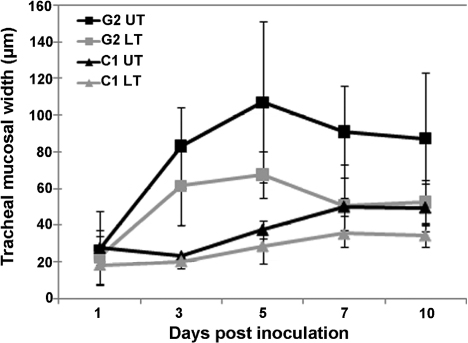
Figure 5. Tracheal mucosal thickness over time for chickens inoculated with N1/08. Error bars represent the standard error of the mean. Group G2 is the group inoculated with N1/08 and group C1 is the negative control group. UT, upper trachea; LT, lower trachea.
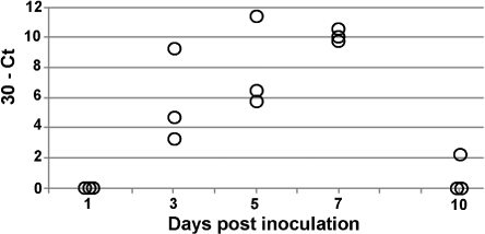
Figure 6. The 30 – Ct values obtained for the tracheal tissues of chickens in group G2. The Ct value indicates at which cycle, during RT-PCR, amplification exceeded a set threshold (Hewson et al., Citation2009). Where values overlapped, horizontal jittering was applied. Each point represents the average 30 – Ct value of the triplicate PCRs performed on each individual tracheal tissue.

Reprints and Corporate Permissions

Please note: Selecting permissions does not provide access to the full text of the article, please see our help page How do I view content?

To request a reprint or corporate permissions for this article, please click on the relevant link below:

Academic Permissions

Please note: Selecting permissions does not provide access to the full text of the article, please see our help page How do I view content?

Obtain permissions instantly via Rightslink by clicking on the button below:

If you are unable to obtain permissions via Rightslink, please complete and submit this Permissions form. For more information, please visit our Permissions help page.