2,007
Views
27
CrossRef citations to date
0
Altmetric
ORIGINAL ARTICLES

Natural infection with highly pathogenic avian influenza virus H5N1 in domestic pigeons (Columba livia) in Egypt

, , , &
Pages 319-324 | Received 09 Jan 2010, Accepted 21 Apr 2014, Published online: 20 Jun 2014

Figures & data

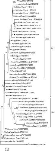
Figure 1. Phylogenetic relationship of pigeon AIV, Egypt 2011 to other AIV H5N1 sequences available in GenBank. The tree was constructed on the basis of the 1594-nucleotide sequence of the HA gene using the neighbour-joining method and the Kimura two-parameter model in MEGA5 (www.megasoftware.net). Bootstrap values of 1000 resamplings as a percentage (>70%) are indicated at key nodes. The virus isolated in this study is marked with a solid square.
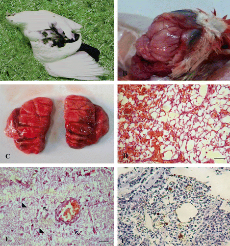
Figure 2. An experimentally infected pigeon at 5 d.p.i. 2a: Severe neurologic signs consisting of torticollis and wing paralysis. 2b: Brain showing severely congested meningeal blood vessels. 2c: Lungs showing oedema and haemorrhage. 2d: Pulmonary haemorrhage represented by occlusion of the alveolar spaces with erythrocytes. Haematoxylin and eosin, bar: 50 µm. 2e: Brain showing vacuolation, spongiosis, degenerated neurons (arrowheads), microglial cell infiltrations (arrows) and perivascular lymphocytic cuffing. Haematoxylin and eosin, bar: 50 µm. 2f: Immunostaining revealed sporadic moderate positive staining of the pneumocytes (asterisk), interstitial tissue and few intravascular mononuclear cells of the lungs (arrows). Immunohistochemistry, bar: 50 µm.
Figure 3. Virus titres in oropharyngeal and cloacal swabs collected from infected and contact pigeons at 3, 5 and 7 d.p.i. The mean value of virus titre (log 10 EID50/ml) in swabs collected from three birds ± standard error.

Reprints and Corporate Permissions

Please note: Selecting permissions does not provide access to the full text of the article, please see our help page How do I view content?

To request a reprint or corporate permissions for this article, please click on the relevant link below:

Academic Permissions

Please note: Selecting permissions does not provide access to the full text of the article, please see our help page How do I view content?

Obtain permissions instantly via Rightslink by clicking on the button below:

If you are unable to obtain permissions via Rightslink, please complete and submit this Permissions form. For more information, please visit our Permissions help page.