Figures & data
Table 1. Mortality in flocks of laying chickens with EPS from which E. coli colonies were subjected to PFGE and MLST genotyping.
Table 2. Results of gross post-mortem examination and bacteriology of dead hens originating from chicken flocks with EPS.
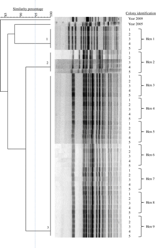
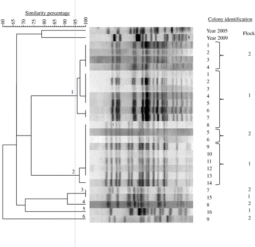
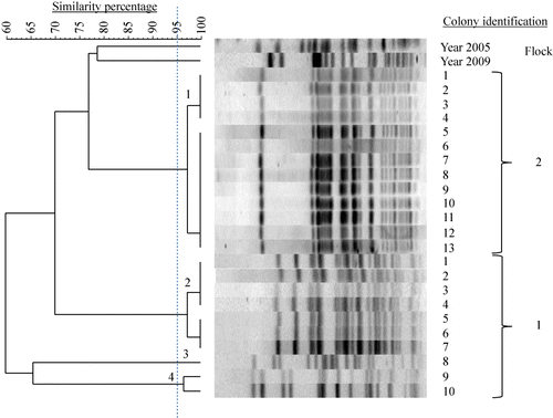
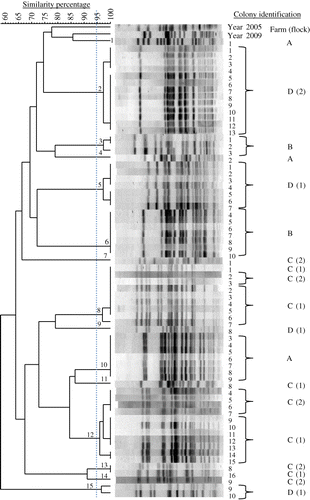
Table 3. Number and prevalence of E. coli PFGE genotypes in chicken flocks with EPS.
Table 4. Target genes, primer name, primer sequence and product size of the seven housekeeping genes used for multilocus sequence typing of E. coli (Wirth et al., Citation2006) isolated from the bone marrow of hens with EPS.
![Figure 1. PFGE dendrogram and corresponding DNA fragment patterns of 45 E. coli colonies obtained from the bone marrow of nine hens with EPS belonging to the same flock (Farm A). Colonies from a single hen (five colonies per hen) were always clonal; however, this was not the case between hens. In this flock, three genotypes were found. Additional colonies of the years 2005 and 2009 from other EPS cases were included.](/cms/asset/a6e61b4a-e916-451c-8a46-038fee79a73e/cavp_a_935291_f0001_oc.jpg)
![Figure 2. PFGE dendrogram and corresponding DNA fragment patterns of 25 E. coli colonies obtained from the bone marrow of single hens with EPS from two successive flocks of Farm C (Flocks 1 and 2). Most colonies of the second flock belonged to genotype 1 or 2 that also dominated in the previous flock. Additional colonies of the years 2005 and 2009 from other EPS cases were included.](/cms/asset/2db6c24a-106c-43f3-a437-d35bb2a9b6f7/cavp_a_935291_f0002_oc.jpg)
![Figure 3. PFGE dendrogram and corresponding DNA fragment patterns of 23 E. coli colonies obtained from the bone marrow of single hens with EPS from two successive flocks of Farm D (Flocks 1 and 2). Genotypes found in the second flock were unrelated to those of the previous one. Additional colonies of the years 2005 and 2009 from other EPS cases were included.](/cms/asset/405b6086-4ee6-44cd-956f-4befe5151c92/cavp_a_935291_f0003_oc.jpg)
![Figure 4. PFGE dendrogram and corresponding DNA fragment patterns of 67 E. coli colonies obtained from the bone marrow of single hens with EPS from six flocks. In total, 15 genotypes were found. Additional colonies of the years 2005 and 2009 from other EPS cases were included.](/cms/asset/91d9e4b4-6691-423f-b0f2-22e4bd2e94e7/cavp_a_935291_f0004_oc.jpg)
![Figure 5. Minimum spanning tree of 63 E. coli colonies obtained from the bone marrow of hens with EPS analysed by MLST. Clustering of MLST profiles was done using a categorical coefficient. Sequence types are displayed as circles. The size of each circle is proportional to the number of E. coli colonies with a particular ST. Inside the circles the coloured area reflects the number of colonies from each collection. Short fat lines connect strains that are different in one single locus. The longer thin line connects double locus variants. Strains that were different in three or more loci are not connected. STs and CCs were assigned using the University of Warwick database. CCs are presented when strains are different in one locus. The shaded grey area around STs denotes types that belong to the same CC, and known CCs are also indicated with a CC and number.](/cms/asset/b8621558-9156-4e16-9c32-345f407aba48/cavp_a_935291_f0005_c.jpg)