2,186
Views
4
CrossRef citations to date
0
Altmetric
ORIGINAL ARTICLES

Efficient Marek's disease virus (MDV) and herpesvirus of turkey infection of the QM7 cell line that does not contain latent MDV genome

, &
Pages 414-419 | Received 03 Dec 2013, Accepted 26 Jul 2014, Published online: 10 Sep 2014

Figures & data

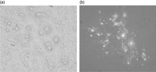
Figure 1. Immunofluoresence and CPE of MDV (652)-infected QM7 foci. QM7 cells were infected with MDV 652 for 7 days in DMEM/F12 with 3% FBS. 1a: CPE of 652/QM7-infected foci. 1b: Infected cells were then fixed and stained with a mixture of monoclonal antibodies H19 (anti-pp38) and 1AN86 (anti-gB). The negative control, QM7 co-cultivated with DEF, stained completely negative.

Table 1. Titration of viral titres for 652/QM7.
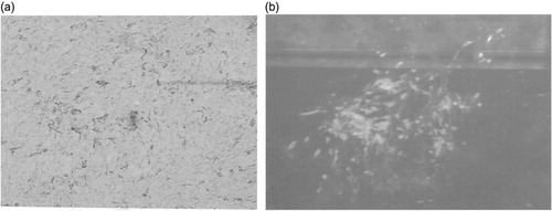
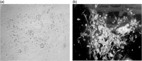
Figure 2. Immunofluoresence and CPE of MDV (652)-transfected QM7 cells. 2a: QM7 cells were transfected with DNA isolated from 652/DEF. Infected foci were seen 7 days post transfection. 2b: Infected cells were then fixed with 80% acetone and stained with a mixture of monoclonal antibodies H19 (anti-pp38) and 1AN86 (anti-gB).
Figure 3. Immunofluoresence and CPE of HVT-infected QM7 foci. QM7 cells were infected with HVT vaccine (MD-Vac; Zoetis, formerly Fort Dodge Animal Health, Fort Dodge, IA, USA) for 7 days in DMEM/F12 with 3% FBS. 3a: CPE of HVT/QM7-infected foci. 3b: Infected cells were then fixed and stained with monoclonal antibody L78. The negative control, QM7, stained completely negative.

Table 2. Different doses of 652/QM7 and 652/DEF that cause in vivo pathogenicity.

Table 3. Virus re-isolation and pathogenicity following in ovo injection.
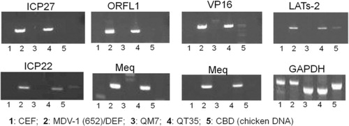
Figure 4. PCR analysis of potential presence of latent MDV genome in avian cells. PCR primers for various MDV gene sequences were used in PCR analysis: ICP4, LAT, ICP22, ICP27, meq, pp38, VP16, ORF L1, and gB. PCR was carried out with 2x JumpStart RED Taq (Sigma). The amplified fragments were resolved by electrophoresis in 2% agarose E-Gels (Invitrogen).

Reprints and Corporate Permissions

Please note: Selecting permissions does not provide access to the full text of the article, please see our help page How do I view content?

To request a reprint or corporate permissions for this article, please click on the relevant link below:

Academic Permissions

Please note: Selecting permissions does not provide access to the full text of the article, please see our help page How do I view content?

Obtain permissions instantly via Rightslink by clicking on the button below:

If you are unable to obtain permissions via Rightslink, please complete and submit this Permissions form. For more information, please visit our Permissions help page.