Figures & data
Figure 1. The spleen, liver and proventricular tissues stained by HE. (A) Numerous small lymphoid tumour cells scattered through the spleen. Scale bar = 25 μm. (B) The liver was infiltrated by the pleomorphic, neoplastic lymphoid tumour cells. Scale bar = 25 μm. (C) The liver was infiltrated by myeloid cells with eosinophilic cytoplasmic granules (myelocytes). Scale bar = 25 μm. (D) A large population of pleomorphic, neoplastic lymphoid tumour cells scattered in the proventricular glands and lamina propria of the proventriculus. Scale bar = 1 mm. (E) A higher magnification of the area indicated by an arrow in D. Scale bar = 50 μm. (F) A higher magnification of the area indicated by an arrow in (E). Two types of tumour cells exist in the same foci in the proventriculus, consisting of myeloid cells with eosinophilic cytoplasmic granules (myelocytes) (black arrow) and lymphoid tumour cells (white arrow). Magnification × 1000.
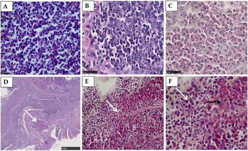
Figure 2. The spleen, liver and proventricular tissues stained by IHC. (A) ALV-J immunoreactivity in the lymphoid tumour cells and parts of the reticular cells in spleen. Magnification × 400. (B) ALV-J immunoreactivity in tumour cells in the liver. Magnification × 400. (C) ALV-J immunoreactivity in tumour cells in the proventricular gland cells. Magnification × 400. (D) MDV immunoreactivity in the lymphoid tumour cells in the spleen. Magnification × 400. (E) MDV immunoreactivity in the lymphoid tumour cells in the liver. Magnification × 400. (F) MDV immunoreactivity in the proventricular gland cells. Magnification × 400. (G) The spleen of non-infected chicken. Magnification × 400. (H) The liver of non-infected chicken. Magnification × 400. (I) The proventriculus of non-infected chicken. Magnification × 400.
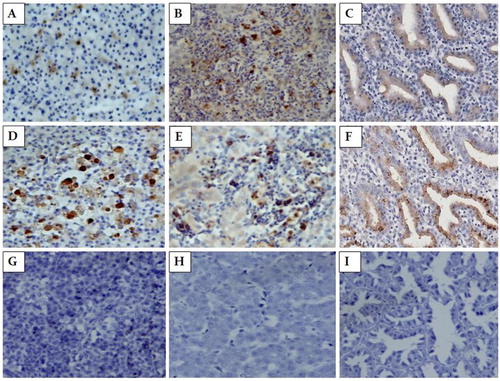
Figure 3. Phylogenetic tree based on the meq and gp85 gene sequences of MDV and ALV-J AN-1 and their reference strains. Unrooted phylogenetic trees were generated using the distance-based neighbour-joining method with the Mega 5 software. (A) The phylogenetic tree for the meq gene from ALV-J AN-1. (B) The phylogenetic tree for the gp85 gene from ALV-J AN-1.
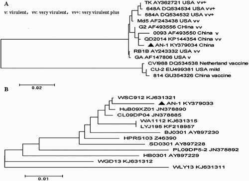
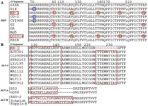
Figure 4. Alignments for the amino acid mutations and deletion sites in the Meq and gp85 protein. (A): Amino acid sequence alignment for the mutation site of the Meq protein from 10 MDVs. (B) Amino acid sequence alignment for the deletion site in the gp85 protein from 10 ALV-Js.