Figures & data
Figure 1. A, B, C: RT-PCR detection of M RNA from buccal swabs of experimentally inoculated canaries. Buccal swabs were sampled at 1, 3, 6, 8, 10 and 14 days pi and RRT-PCR was used to detect viral M RNA. Results are expressed as M RNA copies/ml from swabs sampled from canaries inoculated with the H5N2/chicken virus ((A)), with the H7N1/chicken virus ((B)) and the H5N3/wild mallard virus ((C)). A dotted line represents the approximate limit of detection; samples where virus was not detected are not shown.
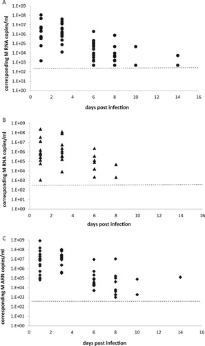
Figure 2. A, B, C: RT-PCR detection of M RNA from fresh and dry faeces. Fresh (cross symbol) and dry (square symbol) faeces were sampled at 1, 3, 6, 8, 10 and 14 days pi and RRT-PCR was used to detect viral M RNA. Results are expressed as M RNA copies/ml from groups of canaries inoculated with the H5N2/chicken virus ((A)), with the H7N1/chicken virus ((B)) and the H5N3/wild mallard virus ((C)). A dotted line represents the approximate limit of detection; samples where virus was not detected were given an arbitrary value of 5.
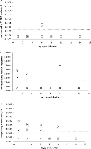
Table 1. Positive samples during the experimental infection of canaries with the H5N2/chicken virus, the H7N1/chicken virus and the H5N3/wild mallard virus.
Figure 3. A, B, C: RT-PCR detection of M RNA in drinking water and water bath samples. Water samples from drinking water (light bars) and bath water (dotted bars) were collected at 1, 3, 6, 8, 10 and 14 days pi and RRT-PCR was used to detect viral M RNA. Results are expressed as M RNA copies/ml from groups of canaries inoculated with the H5N2/chicken virus ((A)), with the H7N1/chicken virus ((B)) and the H5N3/wild mallard virus ((C)). A dotted line represents the approximate limit of detection; samples where virus was not detected were given an arbitrary value of 5.
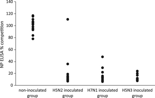
Figure 4. Detection of anti-NP antibodies by competitive NP ELISA. Detection of anti-NP antibodies was performed using a competitive NP ELISA and results are expressed as % of competition. Group 1 is the non-inoculated control group with non-inoculated canaries; group 2: the canaries inoculated with the H5N2/chicken virus; group 3: the canaries inoculated with the H7N1/chicken virus and the group 4: the canaries inoculated with the H5N3/wild mallard virus.