Figures & data
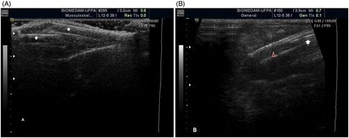
Figure 1. A. Lateral longitudinal view of the femur of a burrowing owl (A. cunicularia). The bone surface of both sides of the cortex (arrows) and the medullary cavity in between can be observed. B. Lateral view of the humerus of a spectacled owl (P. perspicillata). The image shows a cortex in the near field (white arrow) and deep, parallel reverberation artefacts (arrowheads) preventing visualization of deeper structures.
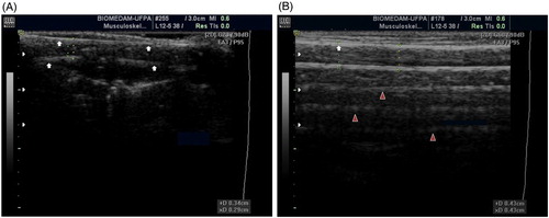
Figure 2. A. Lateromedial view of the tibiotarsus of a hook-billed kite (C. uncinatus). The image shows an incomplete fracture (confirmed after bone dissection) in one of the bone faces while the other face remains intact. B. Lateromedial view of the tibiotarsus of a burrowing owl (A. cunicularia). A metallic object can be observed as an echogenic dot deep to the second face of the bone cortex (arrowhead) confirming the passage of the ultrasound waves through the bone cortex.
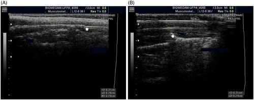
Table 1. Sonographic approaches for each raptor long bone.
Table 2. Mean (±SD) diameter (cm) of the bones from adult raptors of four different species.
Figure 3. A. Lateral view of the humerus of a roadside hawk (R. magnirostris). It is possible to visualize the deep cortex of the diaphysis (white arrow) but not the epiphysis. There is acoustic shadowing preventing visualization of the deeper cortex (arrowhead). B. Craniocaudal view of the femur of a tropical screech owl (M. choliba). The bone diaphysis (arrows) and epiphysis (arrowhead) are shown.
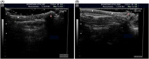
Figure 4. A. Craniocaudal view of the tibiotarsus of a tropical screech owl (M. choliba); the animal arrived at the hospital prostrate and with subcutaneous haemorrhage in the tibial region of the right leg. The image shows a complete oblique fracture with displacement in the mid-diaphysis of the bone (arrows). B. Lateral view of the femur of a barn owl (T. furcata); a metallic pin (white arrow) appears as a linear hyperechoic structure between the near and far cortices of the diaphysis (arrowhead). The image was obtained during the surgical correction of a complete oblique fracture.