Figures & data
Figure 1. Seven-month-old Alagoas curassows (Pauxi mitu) which died of natural infection with Aviadenovirus A. Three birds are shown of four birds which died at the outbreak.
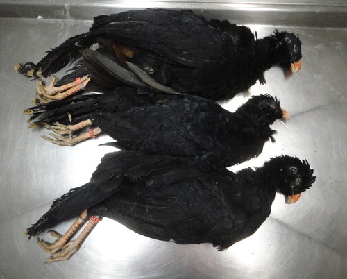
Figure 2. (a) Formalin-fixed trachea from an Alagoas curassow filled with blood and fibrin. (b) Formalin-fixed trachea from an Alagoas curassow with a diphtheritic membrane.
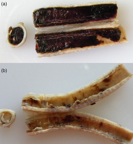
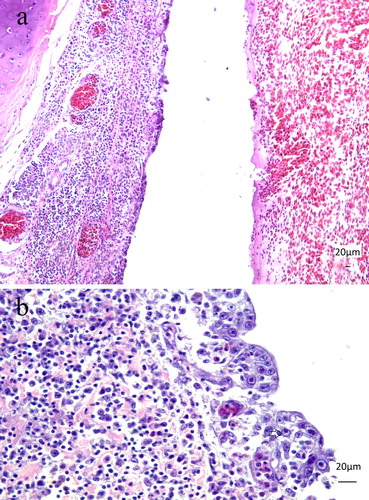
Figure 3. (a) Seven-month-old Alagoas curassow (Pauxi mitu) with tracheitis due to Aviadenovirus. The respiratory epithelium is replaced by a single layer of epithelium, with loss of goblet cells. The lamina propria is expanded by the lymphoplasmacytic infiltration. H&E. Obj. 20. Bar = 20 µm. (b) Seven-month-old Alagoas curassow (P. mitu) with tracheitis by Aviadenovirus. Lymphoplasmacytic tracheitis with epithelial necrosis, loss of goblet cells and basophilic intranuclear inclusion bodies (arrow) were detected. H&E. Obj. 40. Bar = 20 µm.