Figures & data
Table 1. Signalment, clinical signs and clinicopathological findings in coxiellosis in nine psittacine birds, one black-browed barber, and one paradise tanager.
Figure 1. (1) Coxiellosis in a 12-month-old, male budgerigar (case 5). Gross image of the fresh brain tissue, ventral view, showing alternating meningeal hyperaemia (h) and clouding (i; interpreted as inflammation). There is malacia (softening to liquefaction) of the cerebellum, resulting in an irregular surface (*). Photo courtesy of Lauren V Powers, DVM, DABVP. Service Head, Avian and Exotic Pet Service, Carolina Veterinary Specialists,12117 Statesville Road, Huntersville, North Carolina 28078. (2) Coxiellosis in a 12-month-old, male budgerigar (case 5). Haematoxylin and eosin stain, brain, 40×. Meningoencephalitis characterized by increased cellularity of the meninges (black arrow) and the white and grey matter of the brain itself (arrowheads). Bar = 65 µm. Inset – higher magnification showing an infected cell. 400×, bar = 15 µm. (3) Coxiellosis in a male Swainson’s lorikeet (case 6). Haematoxylin and eosin stain, lung, 400×. There is increased cellularity of the interstitium. There are multiple infected cells denoted by the white arrows. Bar = 30 µm. (4) Coxiellosis in a male Swainson’s lorikeet. Gimenez histochemical stain, 400×. The infectious organisms are azurophilic intracytoplasmic inclusions in the infiltrating macrophages. Bar = 30 µm.
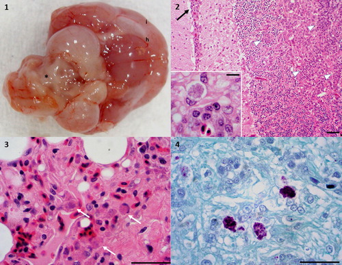
Table 2. Coxiellosis in nine psittacine birds, one black-browed barbet, and one paradise tanager.
Figure 2. Coxiellosis in hyacinth macaw (case 2). Transmission electron micrograph, 14,000×. A macrophage in the spleen contains myriad intracellular bacteria, with peripheralization of the nucleus (N). The membrane of the parasitophorous vacuole (arrowhead) can be seen associated with bacteria (arrow), some of which have a characteristic electron dense core (asterisk). N – host cell nucleus. Bar = 0.5 µm.
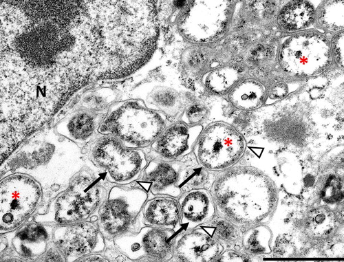
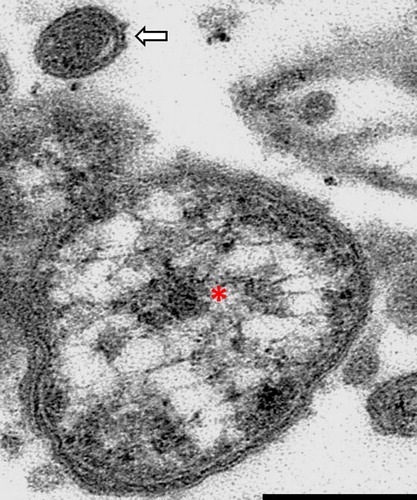
Figure 3. Coxiellosis in hyacinth macaw (case 2). Transmission electron micrograph, 19,000×. An aggregate of bacteria, showing a single large cell variant with electron lucent vacuoles and an electron dense core (asterisk), and a single small cell variant above it (arrow). The small cell variant is almost uniformly electron dense. Note the trilaminar wall of the bacteria. Bar = 0.15 µm.