
Figures & data
Figure 1. Weight (g) of the adult group of cockatiels after infection with PaBV-4 at different ages. Laying females (A0,1, B0,1, C0,1, E0,1 and F0,1) in colour. Arrows mark the timepoint of the first egg of one clutch. Colour online.
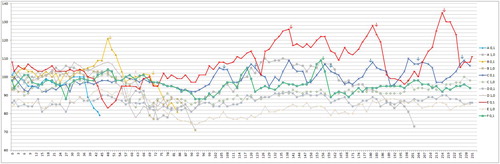
Figure 2. Weight (g) of the juvenile group of cockatiels after infection with PaBV-4 at different ages. Laying females (J3, J4, J7 and J11) as well as the outlier J9 in colour. Arrows mark the timepoint of the first egg of each clutch. Females J3, J4 and J7 were using the same nest, so it was not always possible to know which egg was from which female. The eggs where no assignment to one female was possible are not shown here. Colour online.
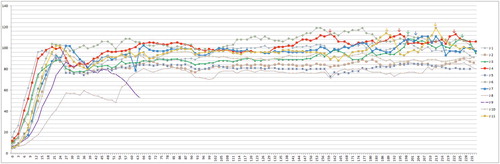
Table 1. Clinical findings of cockatiels after infection with PaBV-4 for the different age groups (values are dpi).
Figure 3. Box plot diagram showing the first detection of anti-PaBV antibodies in cockatiels after infection with PaBV-4 4 in different age groups (juvenile average: 44 dpi, min: 36 dpi, max: 57 dpi; adult average: 54 dpi, min: 36 dpi, max: 71 dpi). The juvenile group seroconverted earlier and more homogenously (P = 0.0502). One outlier in the juvenile group (seroconversion at 99 dpi) was excluded from the diagram.
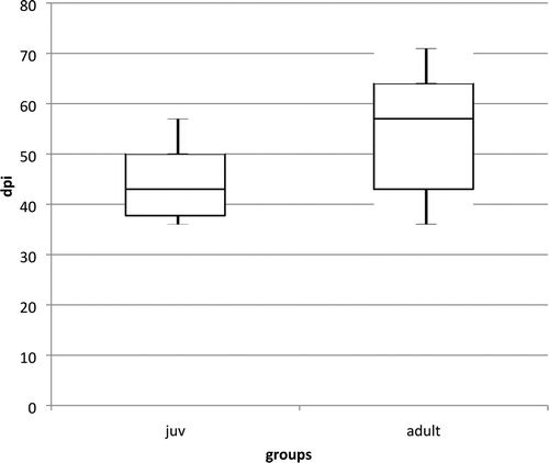
Figure 4. Anti-PaBV-antibody titre of the adult group of cockatiels after infection with PaBV-4 in different age groups in dpi (Y-axis: 1/titre). Colour online.
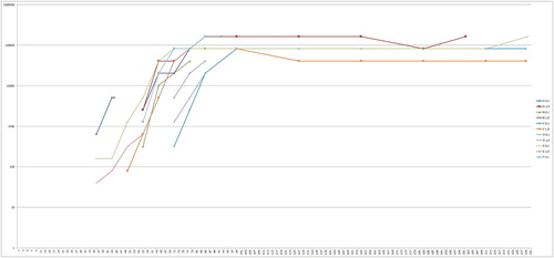
Figure 5. Anti-PaBV-antibody titre of the juvenile group of cockatiels after infection with PaBV-4 in different age groups in dpi (Y-axis: 1/titre). Colour online.
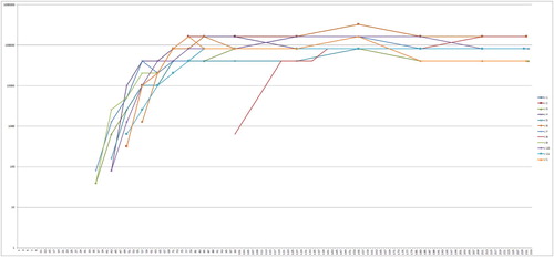
Table 2. Post mortem findings of cockatiels after infection with PaBV-4 in different age groups.
Figure 6. Box plot diagram showing the first detection of PaBV-RNA in combined crop and cloacal swabs of cockatiels after infection with PaBV-4 in different age groups (juvenile average: 31 dpi, min: 27 dpi, max: 36 dpi; adult average: 45 dpi, min: 31 dpi, max: 66 dpi). The juvenile group started shedding earlier and more homogenously, which was highly significant (P = 0.0011). One outlier in the juvenile group (first detection at 52 dpi) was excluded from the diagram.

Table 3. Viral RNA content in the organ samples of cockatiels after infection with PaBV-4 at different ages (Ct-values).