
Figures & data
Figure 1. Experimental West Nile virus (WNV) mean (± standard error of the mean) viraemia profiles for all WNV-inoculated birds in the two age groups and the viraemia profile of the infected sham-inoculated bird in the old group. None of the individual birds developed daily viraemia titres above the solid black line, which represents the peak viraemia titre (105 PFU/ml) deemed sufficient to infect Culex pipiens mosquitoes (Turell et al., Citation2000; Komar et al., Citation2003).
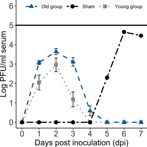
Figure 2. Experimental West Nile virus (WNV) mean (± standard error of the mean) oropharyngeal (A) and cloacal (B) viral shedding profiles for those WNV-inoculated birds in the two age groups that shed virus each day and the oropharyngeal (A) and cloacal (B) viral shedding profiles for the infected sham-inoculated bird in the old group. Numbers correspond to the ratio of WNV-inoculated birds in the young group (bottom) and old group (top) with detectable virus shed each day (and thus included in the mean and standard error of the mean calculations for each day).
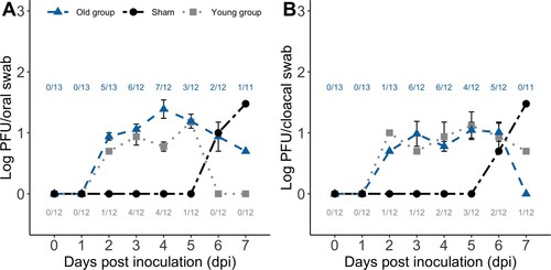
Figure 3. Haematoxylin and eosin (H&E) microscopic lesions and WNV immunohistochemistry (IHC) of experimental WNV-inoculated wild turkeys. (A) Heart; lymphoplasmacytic, histiocytic myocarditis overlay few degenerated cardiomyocytes (old group, 14 dpi, 20×, H&E). (B) Midbrain; lymphoplasmacytic perivascular encephalitis with microgliosis (old group, 14 dpi, 20×, H&E). (C) Caecum; lymphoplasmacytic typhlitis and ganglioneuritis (myenteric plexus) (old group, 14 dpi, 20×, H&E). (D) Proventriculus; serosal lymphoplasmacytic perivascular proventriculitis (old group, 14 dpi, 20×, H&E). (E) Ocular pecten-optic nerve; lymphoplasmacytic pectenitis and neuritis (old group, 14 dpi, 20×, H&E). (F) Testis; lymphoplasmacytic orchitis (old group, 14 dpi, 10×, H&E). (G) Ventriculus; WNV antigen within the cytoplasm of macrophages and lymphocytes in the lamina propria (young group, 14 dpi, 40×, IHC). (H) Lung; few macrophages and lymphocytes in bronchus-associated lymphoid tissue with intracytoplasmic WNV antigen (young group, 14 dpi, 40×, IHC).
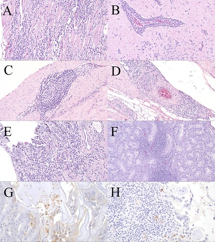
Table 1. Summary of distribution of microscopic lesions and West Nile virus (WNV)-specific immunohistochemical labelling (ratio with microscopic lesions and immunohistochemical labelling and percentage of tissue affected) in tissues of experimentally-inoculated wild turkeys and two sham-inoculated turkeys that became infected.