Figures & data
Table 1. Treatment group descriptions.
Table 2. Number of mallards inoculated with A/AMWI/SC/345/22 H5N1 HPAIV shedding virus and mean titre equivalents based on real-time RT-PCR by dose and day post-challenge.
Figure 1. Titre equivalents of A/American wigeon/SC/000345-01/2022 H5 HPAIV in oral or cloacal swabs by dose group, exposure route and day post-challenge or exposure as determined by quantitative real-time RT-PCR. Thick horizontal bars denote means, thin bars denote standard deviation. The horizontal dotted line is the assay limit of detection. Samples where no virus was detected were given a value of 50% of the assay limit of detection.
Table 3. Distribution of AIV antigen in tissues collected from mallards experimentally infected with A/American Wigeon/South Carolina/000345-001/2022 H5N1 HPAIV. Tissues were collected at 2 and 4 days post-challenge (DPC). The scoring is expressed in the form of bird 1/bird 2 for each DPC. No viral staining was found in the proventriculus, duodenum, caecal tonsils, kidney, and gonads.
Table 4. Titre equivalents (50% egg infectious dose [EID50]) by rRT-PCR of virus in tissues from ducks inoculated with A/American Wigeon/South Carolina/22-000345-001/2022 2 and 4 days post-challenge (DPC).
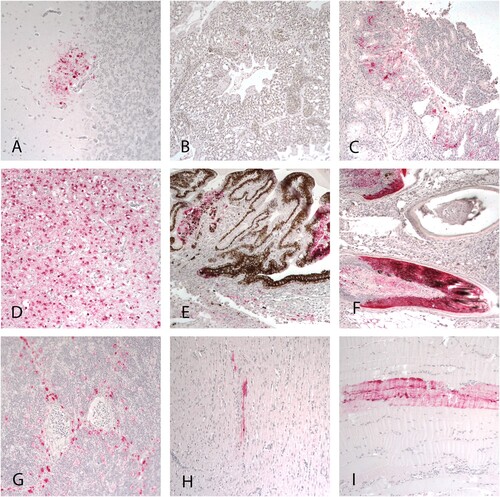
Figure 2. Immunohistochemical staining for AIV antigen in tissues of mallards infected with the A/American Wigeon/SC/000345-01/2022 H5N1 HPAI virus. Virus antigens are stained red. A, B, and C: tissues from ducks at 2 DPC. D-I: tissues collected from ducks at 4 DPC. A. Cerebellum. B. Lung. C. Nasal epithelium. D. Cerebrum. E. Eye ciliary process. F. Feather follicles. G. Thymus. H. Heart. I. Skeletal muscle. Magnification 20×.