
Figures & data
Figure 1. One-day-old chickens showed hepatomegaly and brown (a) to green (b) discolouration of the yolk sac.
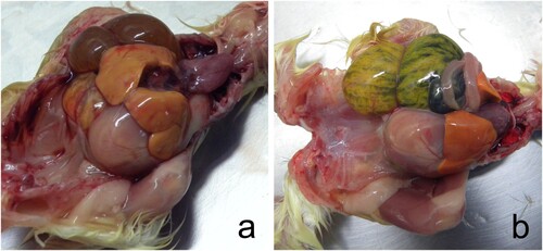
Figure 2. Abnormal egg content consisted of early mortality embryos in different states of decomposition (a, d), middle mortality embryos showing congestion (b, e), and late mortality embryos whose celomatic cavity was open at 21 days of incubation, and the yolk sac was congested (c, d). Orange-brown viscous liquid with a putrid odour was observed in early and middle mortality embryos (a, b, d, e). In late mortality chickens, the coelomic cavity was still open at 21 days of incubation, and the egg yolk was congested with brown content.
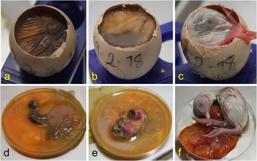
Figure 3. Chicken embryos with late mortality showed brown-orange discolouration of the yolk sac (a, b). The same chicken embryos showing hepatomegaly with congestion (c, d) and multifocal white-spotted foci of necrosis (c) and multifocal coalescent ecchymotic haemorrhages (d).
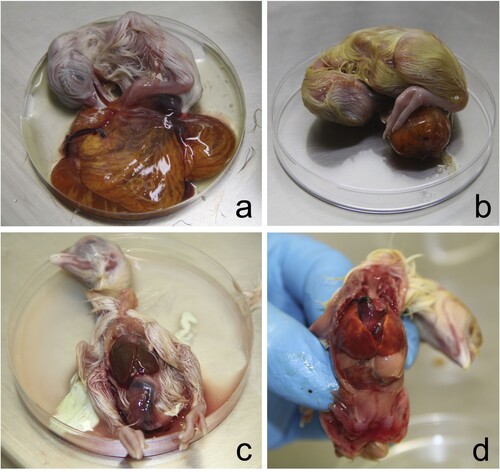
Figure 4. Histological sections of the late mortality embryos showing congestion and interstitial granulocytic infiltration in the bursa of Fabricius (a), kidney (b), liver (c), and the lung (d) (haematoxylin & eosin).
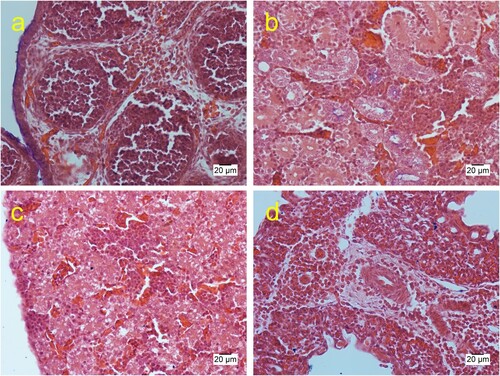
Figure 5. Histological sections of the embryo showing granular deposits around the integument embryo that stain eosinophilic with haematoxylin & eosin (a) and granular dark red with Gram Twort (b, c). Granular deposits with macrophage infiltrations were observed in the integument (a). Detail of the integument showed a feather surrounded by granular deposits stained dark red with Gram Twort (c).
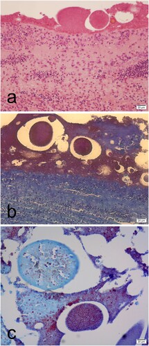
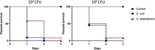
Figure 6. Survival percentages of embryos inoculated with C. testosteroni and E. coli. Groups of 12 embryonated eggs were infected with 102 and 105 C. testosteroni and E. coli. The control group was inoculated with saline solution.