Figures & data
Table 1. List of subfossil and modern Dasycercus specimens examined in this investigation, including study skin specimens examined and their plantar pes morphology.
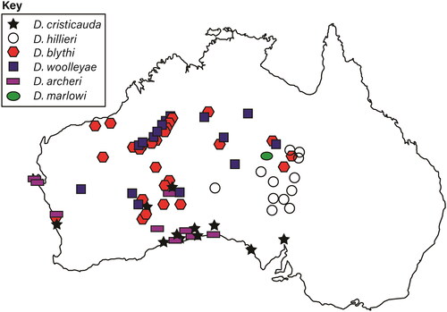
Figure 1. Map of the localities of modern and subfossil Dasycercus specimens used in this investigation. Specimens are labelled using their current museum identification.
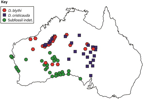
Figure 2. Traditional cranial morphology measurements used in this investigation, adapted from Umbrello (Citation2018). A full list of measurements can be found in Supplementary Data 2. Additionally, illustration of the upper and lower molars of Dasycercus, in occlusal view. Diagram indicates the morphology of the molars, and terminology used in this investigation.
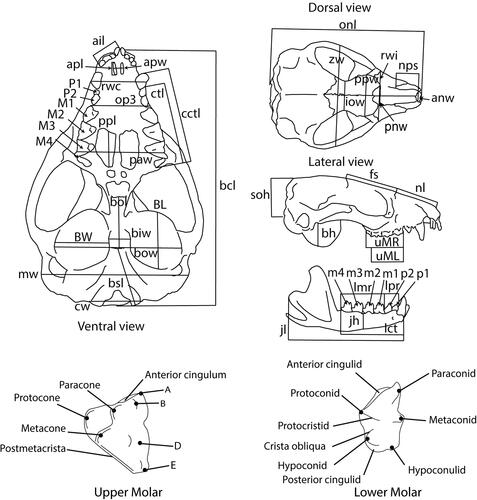
Figure 3. Cranial morphometric analysis of all individuals. A, PCA; B, allometry-corrected PCA; C, LDA; D, allometry-corrected LDA. Males are indicated by filled squares, females by hollow circles, and individuals of indeterminate sex by dashes.
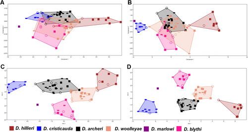
Figure 4. Cranial analysis with females and males separate. A, PCA with females only; B, allometry-corrected PCA with females only; C, PCA with males only; D, allometry-corrected PCA with males only. Not that due to presence of indeterminate material, fewer specimens were available for these analyses.
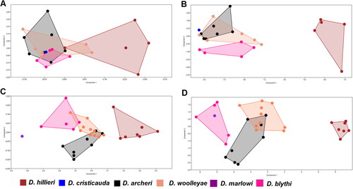
Figure 5. Dental morphometric analysis. A, PCA; B, allometry-corrected PCA; C, LDA; D, allometry-corrected LDA. Males are indicated by filled squares, females by hollow circles, and individuals of indeterminate sex by dashes.
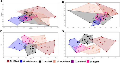
Figure 6. Dental analysis with females and males separate. A, PCA with males only; B, allometry-corrected PCA with males only; C, PCA with females only; D, allometry-corrected PCA with females only. Not that due to presence of indeterminate material, fewer specimens were available for these analyses.
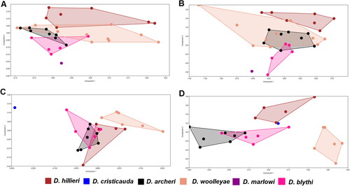
Table 2. P-values of the pairwise one-way PERMANOVA performed on cranial measurements.
Table 3. P-values of the pairwise one-way PERMANOVA performed on allometry corrected cranial measurements.
Table 4. P-values of the pairwise one-way PERMANOVA performed on cranial measurements of male specimens.
Table 5. P-values of the pairwise one-way PERMANOVA performed on allometry corrected cranial measurements of male specimens.
Table 6. P-values of the pairwise one-way PERMANOVA performed on cranial measurements of female specimens.
Table 7. P-values of the pairwise one-way PERMANOVA performed on performed on allometry corrected cranial measurements of female specimens.
Table 8. P-values of the pairwise one-way PERMANOVA performed on dental measurements.
Table 9. P-values of the pairwise one-way PERMANOVA performed on allometry corrected dental measurements.
Figure 7. Phylogenetic relationships of taxa within the genus Dasycercus, and the phylogenetic relationships of Dasycercus within Dasyuromorphia. Bayesian posterior probability support is represented at nodes, as follows: black = BPP ≥ 0.95; dark grey = BPP 0.75–0.94; light grey = BPP 0.5–0.74. Adapted from Kealy & Beck (Citation2017).
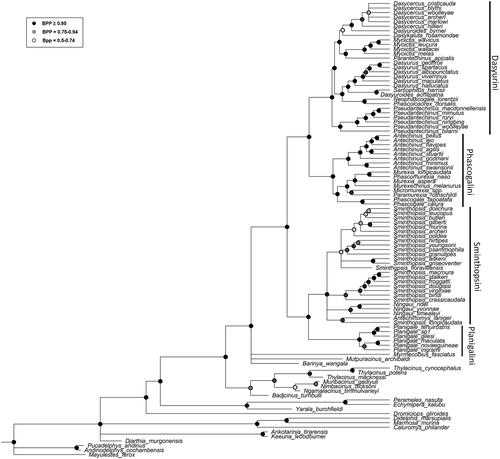
Figure 8. Upper dentition of each taxon of Dasycercus identified in this investigation, specimens are presented in occlusal view. A, D. cristicauda (WAM 67.10.74); B, D. woolleyae (WAM M1513, holotype); C, D. blythi (WAM M1512); D, D. archeri (AMS M2987, holotype); E, D. hillieri (WAM M9670); F, D. marlowi (AMS M8641, holotype). All specimens shown are male.
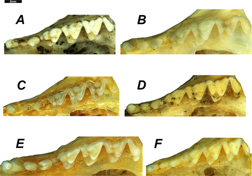
Figure 9. Lower molar rows of each taxon of Dasycercus identified in this investigation (specimens are shown in occlusal view). A, D. hillieri (WAM M9670); B, D. woolleyae (WAM M1513, holotype); C, D. blythi (WAM M1512); D, D. archeri (AMS M2987, holotype); E, D. cristicauda (WAM 67.10.74); F, D. marlowi (AMS M8641, holotype). All specimens shown are male.
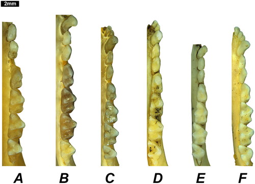
Figure 10. Cranium of each taxon of Dasycercus; D. hillieri (WAM M9670; A, G, M), D. woolleyae (WAM M1513, holotype; B, H, N), D. blythi (WAM M1512; C, I, O), D. archeri (AMS M2987, holotype; D, J, P), D. cristicauda (WAM 67.10.74; E, K, Q), and D. marlowi (AMS M8641, holotype; F, L, R). Specimens are shown in dorsal (A–F), ventral (G–L), and lateral (M–R) views. All specimens shown are male.
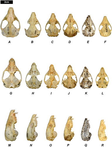
Figure 11. Lateral view of the tails of different Dasycercus taxa, demonstrating the morphology of each. A, D. hillieri (WAM M9670); B, D. blythi (WAM M348); C, D. woolleyae (WAM M1513); D, D. marlowi (AMS M8641); E, D. archeri (AMS M2987). D. cristicauda is not represented due to the poor condition of the holotype specimen. Figures are not to scale.
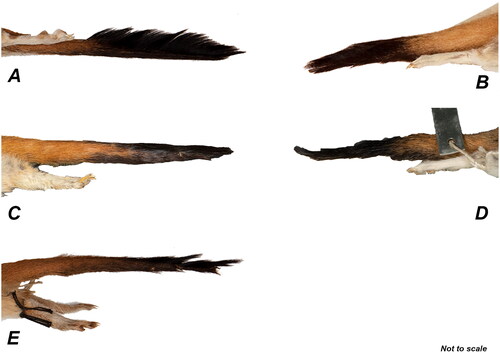
Figure 12. The skin of specimen WAM M8207. This specimen was used to represent D. hillieri. Views are as follows; A, dorsal; B, ventral; C, lateral.
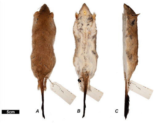
Figure 13. The skin of specimen WAM M348. This specimen was used in this investigation to represent D. blythi. Views are as follows; A, ventral; B, dorsal; C, lateral.
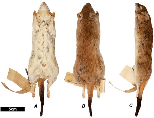
Figure 14. The skin of the holotype specimen of D. woolleyae (WAM M1513), an adult male. Views are as follows: A, ventral; B, dorsal; C, lateral.
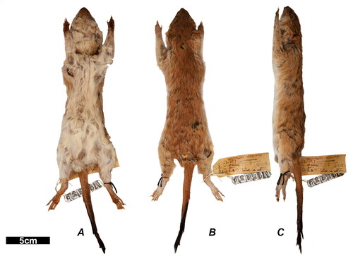
Figure 15. The skin of the holotype specimen of D. archeri (AMS M2987). Views are as follows: A, dorsal; B, ventral; C, lateral.
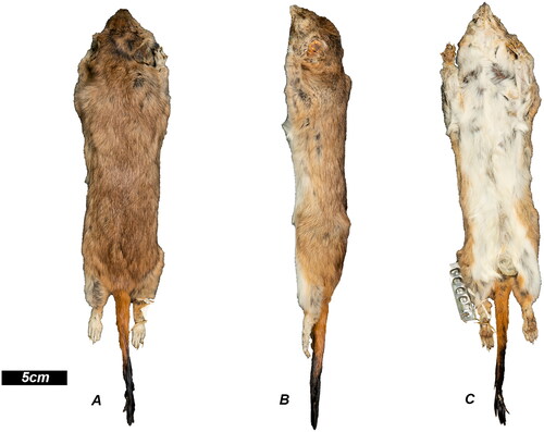
Figure 16. The skin of the holotype specimen of D. marlowi (AMS M8641). Views are as follows: A, dorsal; B, ventral; C, lateral.
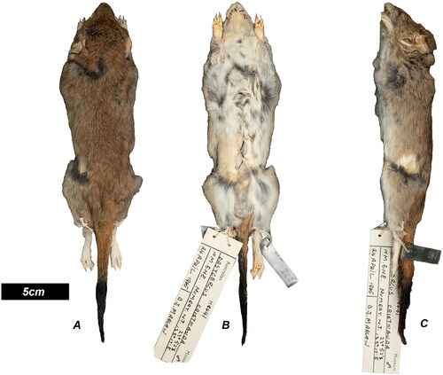
Figure 17. Distribution of Dasycercus material examined in this investigation, specimens have been mapped using taxonomic named assigned in this investigation. Note: material localities have been approximated to protect sensitive sites.