251
Views
1
CrossRef citations to date
0
Altmetric
Forest pathology/Pathologie forestière

Histological investigation of infection processes of Discula destructiva on leaves of Cornus florida

, , , , , & show all
Pages 525-531 | Accepted 06 Oct 2011, Published online: 24 Jan 2012

Figures & data

Figure 1. Experimental wounding tool – a cork stopper embedded with #1 insect pins (16 pins/cm2) – used to abrade adaxial leaf surfaces.
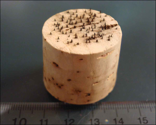
Figure 1. Experimental wounding tool – a cork stopper embedded with #1 insect pins (16 pins/cm2) – used to abrade adaxial leaf surfaces.

Figure 2. Scanning electron micrographs of Cornus florida ‘Cloud 9’ leaves with Discula destructiva conidia. A, Conidia (Con) begin to germinate on the leaf surface underneath the trichome (T) 1 day after inoculation (DAI). B, Germinated conidia with germ tube (GT) on leaf surface 2 DAI. C, Hypha (HY) growing from conidia 3 DAI. Bar = 2 μm.
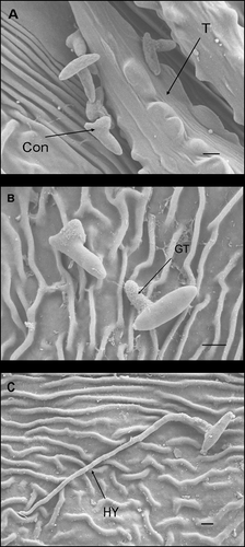
Figure 2. Scanning electron micrographs of Cornus florida ‘Cloud 9’ leaves with Discula destructiva conidia. A, Conidia (Con) begin to germinate on the leaf surface underneath the trichome (T) 1 day after inoculation (DAI). B, Germinated conidia with germ tube (GT) on leaf surface 2 DAI. C, Hypha (HY) growing from conidia 3 DAI. Bar = 2 μm.

Figure 3. Histogram of conidial germination percentage at 1, 2 and 3 days after inoculation (DAI) on potato dextrose agar (PDA) and Cornus florida ‘Cloud 9’, respectively. Sample size n = 100 and bar = 1 SD.
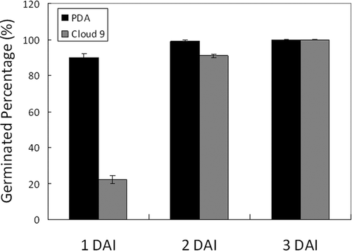
Figure 3. Histogram of conidial germination percentage at 1, 2 and 3 days after inoculation (DAI) on potato dextrose agar (PDA) and Cornus florida ‘Cloud 9’, respectively. Sample size n = 100 and bar = 1 SD.

Figure 4. Scanning electron micrograph of a Discula destructiva hypha (HY) directly penetrating through the Cornus florida ‘Cloud 9’ leaf cuticle (C) and cell wall (CW) into epidermal cells (E) at 3 DAI. Bar = 2 μm.
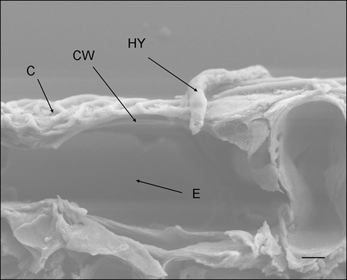
Figure 4. Scanning electron micrograph of a Discula destructiva hypha (HY) directly penetrating through the Cornus florida ‘Cloud 9’ leaf cuticle (C) and cell wall (CW) into epidermal cells (E) at 3 DAI. Bar = 2 μm.

Figure 5. A, B, Light micrographs and scanning electron micrographs, respectively, of cross-sectioned Cornus florida ‘Cloud 9’ leaves inoculated with Discula destructiva conidia at 8 DAI. A, Hypha (HY) is growing intracellularly within epidermal cells (E). Bar = 10 μm. B, Hypha (HY) is growing in epidermal cells. Leaf orientation is adaxial surface up. Bar = 2 μm.
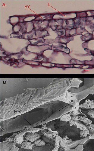
Figure 5. A, B, Light micrographs and scanning electron micrographs, respectively, of cross-sectioned Cornus florida ‘Cloud 9’ leaves inoculated with Discula destructiva conidia at 8 DAI. A, Hypha (HY) is growing intracellularly within epidermal cells (E). Bar = 10 μm. B, Hypha (HY) is growing in epidermal cells. Leaf orientation is adaxial surface up. Bar = 2 μm.

Figure 6. Light micrographs of Cornus florida ‘Cloud 9’ leaves inoculated with Discula destructiva conidia at 16 DAI. A, Diseased infection sites (IS) and healthy tissue (HT). B, Typically diseased, palisade parenchyma (PP) were stained dark red using safranin O. Bar = 20 μm.
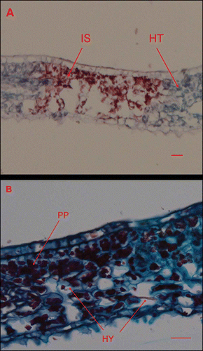
Figure 6. Light micrographs of Cornus florida ‘Cloud 9’ leaves inoculated with Discula destructiva conidia at 16 DAI. A, Diseased infection sites (IS) and healthy tissue (HT). B, Typically diseased, palisade parenchyma (PP) were stained dark red using safranin O. Bar = 20 μm.
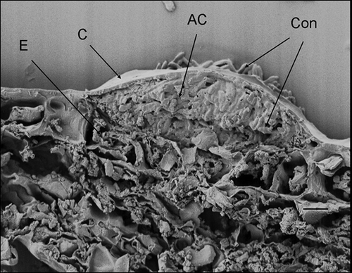
Figure 7. Scanning electron micrograph of Cornus florida ‘Cloud 9’ leaf inoculated with Discula destructiva conidia at 20 DAI. Developing acervulus (AC) with conidia underneath the cuticle and above the epidermal cell layer (E). C, Cuticle; Con, Conidia. Bar = 10 μm.

Figure 7. Scanning electron micrograph of Cornus florida ‘Cloud 9’ leaf inoculated with Discula destructiva conidia at 20 DAI. Developing acervulus (AC) with conidia underneath the cuticle and above the epidermal cell layer (E). C, Cuticle; Con, Conidia. Bar = 10 μm.

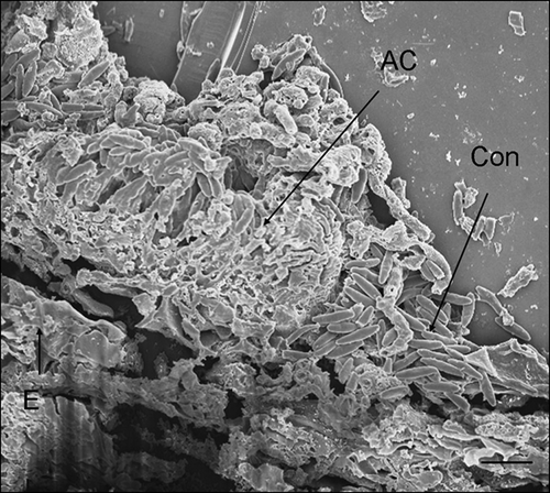
Figure 8. Scanning electron micrograph of Discula destructiva acervulus (AC) fully developed on Cornus florida ‘Cloud 9’ leaf at 24 DAI. Note the crushed epidermal cell layer (E). Con, Conidia. Bar = 10 μm.

Figure 8. Scanning electron micrograph of Discula destructiva acervulus (AC) fully developed on Cornus florida ‘Cloud 9’ leaf at 24 DAI. Note the crushed epidermal cell layer (E). Con, Conidia. Bar = 10 μm.

Reprints and Corporate Permissions

Please note: Selecting permissions does not provide access to the full text of the article, please see our help page How do I view content?

To request a reprint or corporate permissions for this article, please click on the relevant link below:

Academic Permissions

Please note: Selecting permissions does not provide access to the full text of the article, please see our help page How do I view content?

Obtain permissions instantly via Rightslink by clicking on the button below:

If you are unable to obtain permissions via Rightslink, please complete and submit this Permissions form. For more information, please visit our Permissions help page.