Figures & data
Fig. 1. Response of lentil cultivar ‘CDC Robin’ with partial resistance to race 1 isolates 10 days post-inoculation with Colletotrichum truncatum isolates CT-15, CT-21 and CT-35 (all race 1), and CT-20, CT-30 and CT-34 (all race 0).
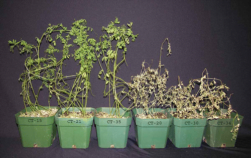
Fig. 2. Conidial germination (per cent) and appressorium formation (as per cent of conidia) of six isolates of Colletotrichum truncatum belonging to two races on detached leaflets of lentil cultivars ‘Eston’ and ‘CDC Robin’ 12 h post-inoculation.
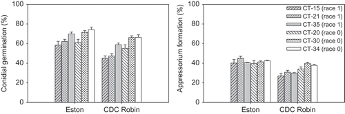
Fig. 3. Stages in the infection process of Colletrotrichum truncatum on lentil leaflets. Conidia germinate and produce appressoria (a). Upon penetration of the host tissue (b), an infection vesicle is produced (arrow) and (c) elongates into a primary hypha (ph). As the primary hypha continues to grow in the epidermal host cell it becomes branched and lobed (d). By the time secondary hyphae (sh) grow rapidly through the surrounding host tissue, the primary hypha has filled the host epidermal cell (e). Appressoria are denoted with an *. Bars = 10 μm.
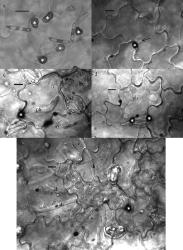
Fig. 4. Penetration (as per cent of appressoria) of six isolates of Colletotrichum truncatum belonging to two races into detached leaflets of lentil cultivars ‘Eston’ and ‘CDC Robin’.
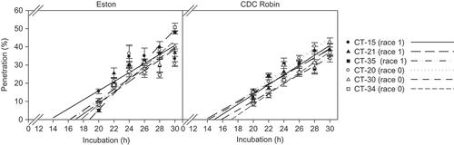
Fig. 5. Size of primary hyphae (assessed as per cent epidermal cell area occupied by primary hyphae) of six isolates of Colletotrichum truncatum belonging to two races 48 h and 60 h post-inoculation in detached leaflets of lentil cultivars ‘Eston’ and ‘CDC Robin’.
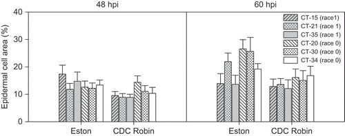
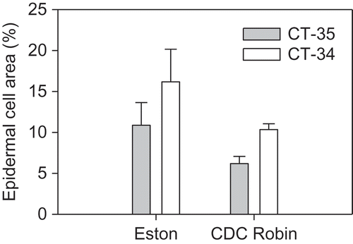
Fig. 6. Size of primary hyphae (assessed as per cent epidermal cell area occupied by primary hyphae) of Colletotrichum truncatum isolates CT-34 (race 0) and CT-35 (race 1) 48 h post-inoculation in attached leaflets of cultivars ‘Eston’ and ‘CDC Robin’.