300
Views
3
CrossRef citations to date
0
Altmetric
Virology/Virologie

Ultrastructural effects of infection caused by Tobacco rattle virus transmitted by Trichodorus primitivus in potato and tobacco tissues

, , &
Pages 126-138 | Accepted 06 Feb 2012, Published online: 27 Apr 2012

Figures & data

Fig. 1. a, External surface necrosis (white arrows) and leaflet deformation (black arrows) on tobacco ‘Samsun’ 4 weeks after plants were introduced into soil with nematodes. b, External stem (long arrows) and leaf petioles (short arrows) with necrosis 4 weeks after TRV PSG was transferred by Trichodorus primitivus into tobacco ‘Samsun’. c, Ectoparasitic nematodes Trichodorus primitivus (Nm) feeding on roots of potato (R) ‘Glada’ 3 weeks after the plant was placed in infested soil. Bar = 100 μm. d, Cross-section of the body (asterisk) of the nematode near root hairs (RH) of tobacco. Bar = 1 μm.
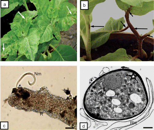
Fig. 1. a, External surface necrosis (white arrows) and leaflet deformation (black arrows) on tobacco ‘Samsun’ 4 weeks after plants were introduced into soil with nematodes. b, External stem (long arrows) and leaf petioles (short arrows) with necrosis 4 weeks after TRV PSG was transferred by Trichodorus primitivus into tobacco ‘Samsun’. c, Ectoparasitic nematodes Trichodorus primitivus (Nm) feeding on roots of potato (R) ‘Glada’ 3 weeks after the plant was placed in infested soil. Bar = 100 μm. d, Cross-section of the body (asterisk) of the nematode near root hairs (RH) of tobacco. Bar = 1 μm.

Fig. 2. a, Necrosis of rhizodermis (long arrows) and fracture of cell walls (CW, black asterisk) in primary cortex. Necrosis of primary cortex parenchyma cells (PCP, white asterisks) of tobacco root can be seen. X = xylem tracheary element. Bar = 50 μm. b, Primary cortex cells (PCP) with necrosis of phloem parenchyma (white arrows) and primary cortex (dark arrows) of root of potato ‘Glada’. Ph = phloem, X = xylem tracheary element. Bar = 50 μm. c, Necrosis of phloem parenchymal cells (asterisks), primary cortex parenchyma (PCP, white arrow) and pith parenchymal cells (dark arrow) of tobacco stem. X = xylem tracheary element. Bar = 50 μm. d, Necrosis of palisade mesophyll cells (Pme, asterisks) of tobacco leaf blade. Sme = spongy mesophyll, VB = vascular bundle. Bar = 50 μm.
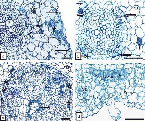
Fig. 2. a, Necrosis of rhizodermis (long arrows) and fracture of cell walls (CW, black asterisk) in primary cortex. Necrosis of primary cortex parenchyma cells (PCP, white asterisks) of tobacco root can be seen. X = xylem tracheary element. Bar = 50 μm. b, Primary cortex cells (PCP) with necrosis of phloem parenchyma (white arrows) and primary cortex (dark arrows) of root of potato ‘Glada’. Ph = phloem, X = xylem tracheary element. Bar = 50 μm. c, Necrosis of phloem parenchymal cells (asterisks), primary cortex parenchyma (PCP, white arrow) and pith parenchymal cells (dark arrow) of tobacco stem. X = xylem tracheary element. Bar = 50 μm. d, Necrosis of palisade mesophyll cells (Pme, asterisks) of tobacco leaf blade. Sme = spongy mesophyll, VB = vascular bundle. Bar = 50 μm.

Fig. 3. a, Complete TRV particles (VP, arrows) of two lengths (L and S) in tobacco rhizodermis cell (Rh). Bar = 0.2 μm. b, Complete TRV PSG particles (VP) of two lengths (arrows) near cells wall (CW) of primary cortex parenchymal cell (PCP) of tobacco root. Cell wall has clearly loosened structure. M = mitochondria. Bar = 0.2 μm. c, Vast necrosis in rhizodermis (Rh) and primary cortex parenchyma (PCP) of tobacco root. Tissue system disrupted by strong deformations of cell walls (asterisks, CW). Bar = 1 μm. d, Complete TRV particles (VP) at cross-section (arrow) in an area isolated by deformed cell walls (CW) of rhizodermis. Bar = 0.1 μm.
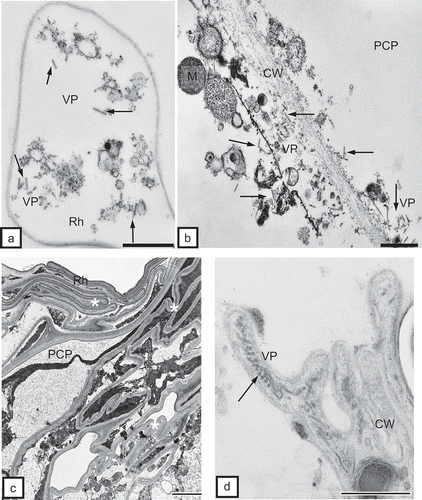
Fig. 3. a, Complete TRV particles (VP, arrows) of two lengths (L and S) in tobacco rhizodermis cell (Rh). Bar = 0.2 μm. b, Complete TRV PSG particles (VP) of two lengths (arrows) near cells wall (CW) of primary cortex parenchymal cell (PCP) of tobacco root. Cell wall has clearly loosened structure. M = mitochondria. Bar = 0.2 μm. c, Vast necrosis in rhizodermis (Rh) and primary cortex parenchyma (PCP) of tobacco root. Tissue system disrupted by strong deformations of cell walls (asterisks, CW). Bar = 1 μm. d, Complete TRV particles (VP) at cross-section (arrow) in an area isolated by deformed cell walls (CW) of rhizodermis. Bar = 0.1 μm.

Fig. 4. a, Complete TRV PSG particles (VP) of two lengths near the Golgi apparatuses (GA) and numerous vesicles (vs) in the protoplast of the primary cortex parenchymal cell (PCP) of potato root. CW = cell wall. Bar = 0.2 μm. b, Dispersed incomplete TRV particles (VP) near Golgi apparatus (GA) in phloem fibre (Pf) of tobacco root. CW = cell wall. Bar = 0.5 μm. c, Complete TRV PSG particles (arrow) in phloem parenchymal cell (PP) of potato ‘Glada’ root. The marked fragment is magnified in CC = companion cell, N = nucleus, Pf = phloem fibres, SE = sieve element. Bar = 1 μm. d, Magnified fragment from CW = cell wall, GA = Golgi apparatus, M = mitochondria,N = nucleus, VP = virus particles. Bar = 0.2 μm.
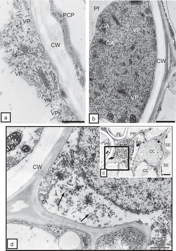
Fig. 4. a, Complete TRV PSG particles (VP) of two lengths near the Golgi apparatuses (GA) and numerous vesicles (vs) in the protoplast of the primary cortex parenchymal cell (PCP) of potato root. CW = cell wall. Bar = 0.2 μm. b, Dispersed incomplete TRV particles (VP) near Golgi apparatus (GA) in phloem fibre (Pf) of tobacco root. CW = cell wall. Bar = 0.5 μm. c, Complete TRV PSG particles (arrow) in phloem parenchymal cell (PP) of potato ‘Glada’ root. The marked fragment is magnified in Fig. 4d. CC = companion cell, N = nucleus, Pf = phloem fibres, SE = sieve element. Bar = 1 μm. d, Magnified fragment from Fig. 4c. CW = cell wall, GA = Golgi apparatus, M = mitochondria,N = nucleus, VP = virus particles. Bar = 0.2 μm.

Fig. 5. a, Incomplete TRV PSG particles (VP, arrows) in a companion cell (CC) of tobacco root. CW = cell wall, PD = plasmodesmata, SE = sieve element. Bar = 0.2 μm. b, Incomplete TRV PSG particles (VP, arrows) in a mature tracheary element of xylem (X) of tobacco root. XP = xylem parenchyma. Bar = 0.2 μm. c, Arrows indicate complete virus particles in xylem tracheary element (X). M = mitochondria. Bar = 0.2 μm. d, Incomplete TRV PSG particles (VP, arrows) in a mature tracheary element of xylem (X) of potato root. XP = xylem parenchyma. Bar = 0.5 μm. e, Complete (arrows) and incomplete (asterisks) TRV particles in xylem parenchymal cells (XP) of potato root. CW = cell wall. Bar = 200 nm.
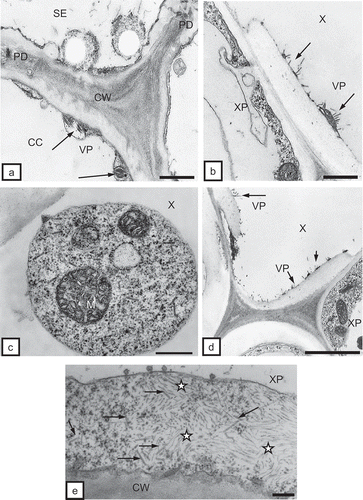
Fig. 5. a, Incomplete TRV PSG particles (VP, arrows) in a companion cell (CC) of tobacco root. CW = cell wall, PD = plasmodesmata, SE = sieve element. Bar = 0.2 μm. b, Incomplete TRV PSG particles (VP, arrows) in a mature tracheary element of xylem (X) of tobacco root. XP = xylem parenchyma. Bar = 0.2 μm. c, Arrows indicate complete virus particles in xylem tracheary element (X). M = mitochondria. Bar = 0.2 μm. d, Incomplete TRV PSG particles (VP, arrows) in a mature tracheary element of xylem (X) of potato root. XP = xylem parenchyma. Bar = 0.5 μm. e, Complete (arrows) and incomplete (asterisks) TRV particles in xylem parenchymal cells (XP) of potato root. CW = cell wall. Bar = 200 nm.

Fig. 6. a, Inclusions of incomplete TRV PSG particles (VP, arrows) in mesophyll cells of tobacco leaves. Protoplasts in a distance to cell wall (asterisks, CW). Ch = chloroplast. Bar = 1 μm. b, Incomplete TRV particles (VP) in the intercellular space (Int) of palisade mesophyll (Pme). Ch = chloroplast, CW = cell wall. Bar = 0.5 μm. c, Fractured cell wall (CW) between cells of palisade mesophyll (Pme). Numerous incomplete TRV particles (VP) of two lengths are present. Changed chloroplast (Ch) structure. ER = endoplasmic reticulum. Bar = 0.5 μm.
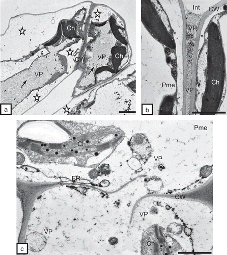
Fig. 6. a, Inclusions of incomplete TRV PSG particles (VP, arrows) in mesophyll cells of tobacco leaves. Protoplasts in a distance to cell wall (asterisks, CW). Ch = chloroplast. Bar = 1 μm. b, Incomplete TRV particles (VP) in the intercellular space (Int) of palisade mesophyll (Pme). Ch = chloroplast, CW = cell wall. Bar = 0.5 μm. c, Fractured cell wall (CW) between cells of palisade mesophyll (Pme). Numerous incomplete TRV particles (VP) of two lengths are present. Changed chloroplast (Ch) structure. ER = endoplasmic reticulum. Bar = 0.5 μm.

Fig. 7. a, Complete TRV PSG particles (arrows) of two lengths near chloroplasts (Ch) in a potato mesophyll cell. Absence of visible double membrane around chloroplasts (asterisks). Bar = 0.2 μm. b, Regular inclusion of TRV particles (VP) made of complete (dark arrow) and incomplete (white arrow) particles. Complete particles present also inside vacuoles (asterisks) of tobacco mesophyll cells. CW = cell wall, ER = endoplasmic reticulum. Bar = 200 nm. c, Incomplete TRV PSG particles (VP) in xylem parenchyma (XP) of tobacco leaf. Particles are present in the apoplast (asterisks) between the protoplast and the cell wall (CW) and in cytoplasm and vacuoles (V). Ch = chloroplast, N = nucleus, X = xylem tracheary element. Bar = 1μm. d, Complete TRV particles (VP) of two lengths inside the xylem (X) tracheary element of potato leaf. CW = cell wall. Bar = 0.5 μm.
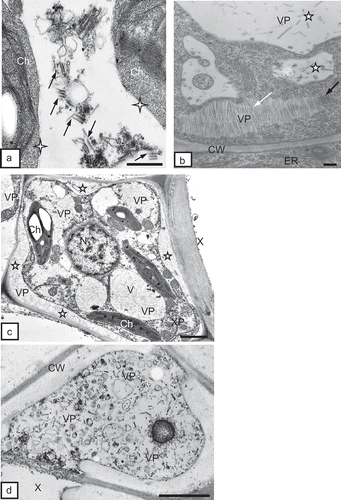
Fig. 7. a, Complete TRV PSG particles (arrows) of two lengths near chloroplasts (Ch) in a potato mesophyll cell. Absence of visible double membrane around chloroplasts (asterisks). Bar = 0.2 μm. b, Regular inclusion of TRV particles (VP) made of complete (dark arrow) and incomplete (white arrow) particles. Complete particles present also inside vacuoles (asterisks) of tobacco mesophyll cells. CW = cell wall, ER = endoplasmic reticulum. Bar = 200 nm. c, Incomplete TRV PSG particles (VP) in xylem parenchyma (XP) of tobacco leaf. Particles are present in the apoplast (asterisks) between the protoplast and the cell wall (CW) and in cytoplasm and vacuoles (V). Ch = chloroplast, N = nucleus, X = xylem tracheary element. Bar = 1μm. d, Complete TRV particles (VP) of two lengths inside the xylem (X) tracheary element of potato leaf. CW = cell wall. Bar = 0.5 μm.

Reprints and Corporate Permissions

Please note: Selecting permissions does not provide access to the full text of the article, please see our help page How do I view content?

To request a reprint or corporate permissions for this article, please click on the relevant link below:

Academic Permissions

Please note: Selecting permissions does not provide access to the full text of the article, please see our help page How do I view content?

Obtain permissions instantly via Rightslink by clicking on the button below:

If you are unable to obtain permissions via Rightslink, please complete and submit this Permissions form. For more information, please visit our Permissions help page.