1,334
Views
74
CrossRef citations to date
0
Altmetric
Disease control/Moyens de lutte

Antagonism of the endophytic insect pathogenic fungus Metarhizium robertsii against the bean plant pathogen Fusarium solani f. sp. phaseoli

&
Pages 288-293 | Received 15 Apr 2013, Accepted 25 Jun 2013, Published online: 29 Jul 2013

Figures & data

Fig. 1. Images of dual plate cultures of Metarhizium robertsii (M.r) and Fusarium solani (F.s) and biocontrol of F. solani by M. robertsii on bean root. (a) Dual plate assays showing antagonistic activity of M. robertsii on F. solani on day 7 and 14. In each photo, a zone of F. solani clearing is evident at the colony interface. (b) Images of bean root rot caused by F. solani after four weeks. Roots were obtained from control plants, F. solani treatment and M. robertsii + F. solani treatments.
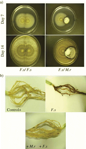
Fig. 1. Images of dual plate cultures of Metarhizium robertsii (M.r) and Fusarium solani (F.s) and biocontrol of F. solani by M. robertsii on bean root. (a) Dual plate assays showing antagonistic activity of M. robertsii on F. solani on day 7 and 14. In each photo, a zone of F. solani clearing is evident at the colony interface. (b) Images of bean root rot caused by F. solani after four weeks. Roots were obtained from control plants, F. solani treatment and M. robertsii + F. solani treatments.

Fig. 2. Inhibition of Fusarium solani in Metarhizium robertsii cell-free culture extracts. (a) Germination of F. solani conidia in M. robertsii cell-free culture extract. Fusarium solani conidia were suspended in different concentrations (100%, 90%, 50% and 10%) of 4-day-old and 10-day-old M. robertsii cell-free culture filtrates. Con = control, no cell-free extract. Germination of 100 F. solani conidia was counted (N = 4) after 24 h (light bars) and 48 h (dark bars). Each bar represents mean % germination ± S. E. (b) Fusarium solani biomass yield in M. robertsii cell-free culture filtrate (autoclaved – light bars and non-autoclaved – dark bars) after four days of growth. Data represents the mean ± S.E. of dry weights of F. solani grown under different concentrations (100%, 90% and 50%) of M. robertsii autoclaved and not autoclaved cell-free extract and the control (Con = control, no cell-free extract).
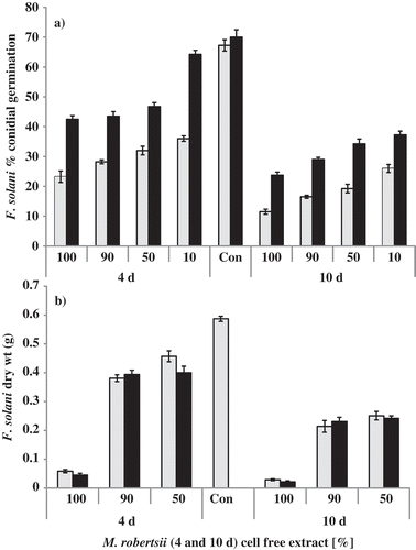
Fig. 2. Inhibition of Fusarium solani in Metarhizium robertsii cell-free culture extracts. (a) Germination of F. solani conidia in M. robertsii cell-free culture extract. Fusarium solani conidia were suspended in different concentrations (100%, 90%, 50% and 10%) of 4-day-old and 10-day-old M. robertsii cell-free culture filtrates. Con = control, no cell-free extract. Germination of 100 F. solani conidia was counted (N = 4) after 24 h (light bars) and 48 h (dark bars). Each bar represents mean % germination ± S. E. (b) Fusarium solani biomass yield in M. robertsii cell-free culture filtrate (autoclaved – light bars and non-autoclaved – dark bars) after four days of growth. Data represents the mean ± S.E. of dry weights of F. solani grown under different concentrations (100%, 90% and 50%) of M. robertsii autoclaved and not autoclaved cell-free extract and the control (Con = control, no cell-free extract).

Table 1.  The effect of M. robertsii (M.r) as an antagonist of Fusarium solani (F.s) infection of bean plants

Reprints and Corporate Permissions

Please note: Selecting permissions does not provide access to the full text of the article, please see our help page How do I view content?

To request a reprint or corporate permissions for this article, please click on the relevant link below:

Academic Permissions

Please note: Selecting permissions does not provide access to the full text of the article, please see our help page How do I view content?

Obtain permissions instantly via Rightslink by clicking on the button below:

If you are unable to obtain permissions via Rightslink, please complete and submit this Permissions form. For more information, please visit our Permissions help page.