Figures & data
Fig. 1. Inhibition of growth of Monilinia fructicola isolates on PDA amended with fenbuconazole deposited by spiral gradient dilution (left) and a control dish without fungicide (right). The concentration of the fungicide decreases toward the edge of the dish (see text for details).
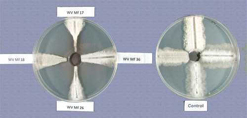
Fig. 2. Effective concentrations for 50% inhibition (EC50) of mycelial growth of Monilinia fructicola isolates by fenbuconazole. Bars in columns represent standard error of the means.
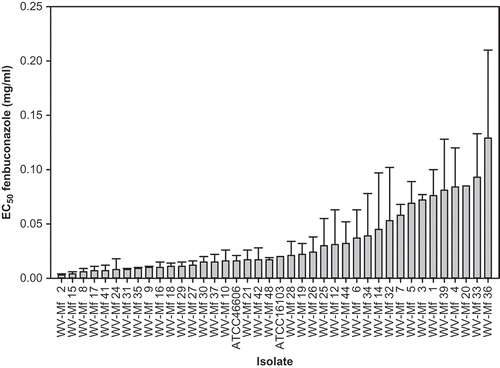
Fig. 3. An example of vegetative compatibility groups (VCG) of Monilinia fructicola isolates. Numbers correspond to the isolate number e.g. 1 = WV-Mf 1 and designation B following the location (AFRS) corresponds to one of the two (A and B) single-spore clones of the same isolates, and only interactions between B clones are presented. Note the strong barrage formation between isolates (arrows). There was no interaction between clones A and B of the isolates in control treatments.
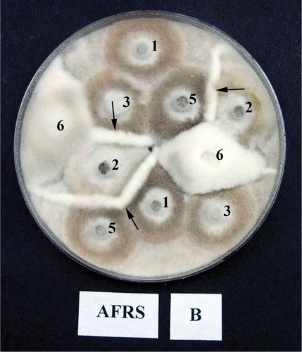
Table 1. List of Monilinia fructicola isolates used in this study and visual appearance of the colonies on agar media
Table 2. Growth rate per day (mm) on PDA medium of Monilinia fructicola isolates at three temperatures
Fig. 4. (Continued.)
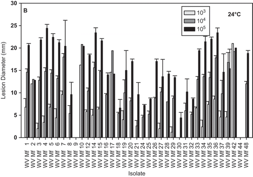
Fig. 5. Brown rot lesion development on wound-inoculated nectarine fruit with mycelium of different Monilinia fructicola isolates incubated at: A, 4 °C for 17 days; and B, 24 °C for 2 days. * Lesions developed on nectarines inoculated with isolate WV-Mf 38 only after additional incubation for three days.
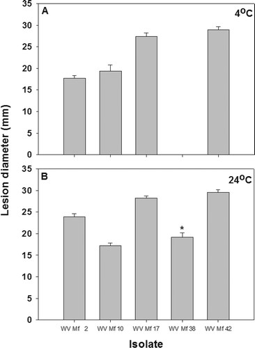
Fig. 6. Regression analysis of isolate growth on PDA against lesion diameter on nectarines inoculated with three concentrations (103, 104 and 105 conidial mL−1) of Monilinia fructicola conidia and incubated at 24 °C for 2 days.
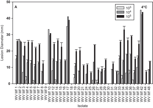
Fig. 4. Brown rot lesion development on wound-inoculated nectarine fruit with 25 μL of conidial suspensions of different Monilinia fructicola isolates at 103, 104 and 105conidia mL−1 and incubated at: A, 4 °C for 17 days and B, 24 °C for 2 days.