161
Views
6
CrossRef citations to date
0
Altmetric
Epidemiology/Épidémiologie

Comparison of macro-morphological and physiological methods for Monilinia species identification in Paraná State, Brazil

, , &
Pages 38-47 | Accepted 06 Nov 2013, Published online: 20 Mar 2014

Figures & data

Fig. 1. Macro-morphological features for identification of Monilinia spp. isolates. Colonies after 10 days of incubation at 22 ºC and 12 h light regime on acidified potato dextrose agar. A, Monilinia laxa – isolate number 15. B, Monilinia fructicola – isolate number 50.C, Monilinia fructigena – isolate number 152. (1) upper colony surface, (2) underside of colony.
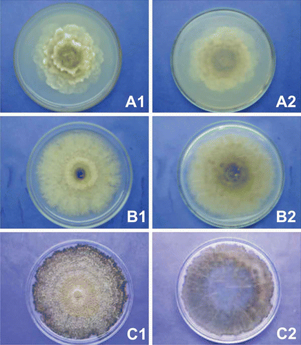
Fig. 1. Macro-morphological features for identification of Monilinia spp. isolates. Colonies after 10 days of incubation at 22 ºC and 12 h light regime on acidified potato dextrose agar. A, Monilinia laxa – isolate number 15. B, Monilinia fructicola – isolate number 50.C, Monilinia fructigena – isolate number 152. (1) upper colony surface, (2) underside of colony.

Table 1. Assignment of synoptic key letters to Monilinia isolates identification based in macro-morphological features.

Fig. 2. Growth interval and optimum growth temperature for Monilinia spp. separated in groups based on macro-morphological characteristics and on the cubic model adjusted. Mycelial growth was assessed after 4 days of growth at 10 °C, 15 ° C, 20 ° C, and 25 ° C with 12 h light regime. Arabic and Roman numerals indicate isolates and macro-morphological groups, respectively.
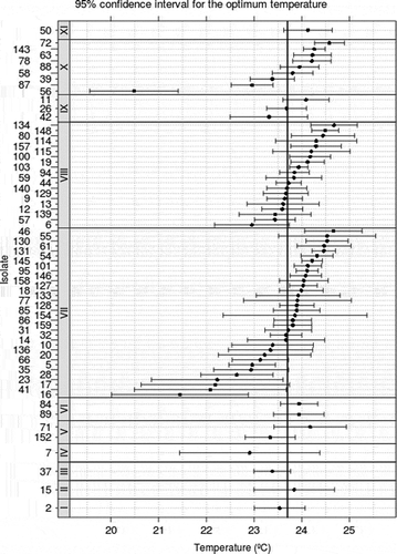
Fig. 2. Growth interval and optimum growth temperature for Monilinia spp. separated in groups based on macro-morphological characteristics and on the cubic model adjusted. Mycelial growth was assessed after 4 days of growth at 10 °C, 15 ° C, 20 ° C, and 25 ° C with 12 h light regime. Arabic and Roman numerals indicate isolates and macro-morphological groups, respectively.
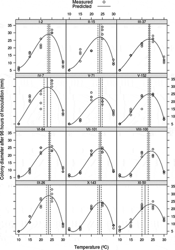
Fig. 3. Growth interval and optimum growth temperature for Monilinia spp. separated by isolates and based on the cubic model adjusted. Mycelial growth was assessed after 4 days of growth at 10 ° C, 15 ° C, 20 ° C, and 25 ° C with 12 h light regime. Arabic and Roman numerals indicate isolates and macro-morphological groups, respectively.

Fig. 3. Growth interval and optimum growth temperature for Monilinia spp. separated by isolates and based on the cubic model adjusted. Mycelial growth was assessed after 4 days of growth at 10 ° C, 15 ° C, 20 ° C, and 25 ° C with 12 h light regime. Arabic and Roman numerals indicate isolates and macro-morphological groups, respectively.

Fig. 4. Pathogenicity of Monilinia spp. isolate groups. Lesion growth was assessed after 4 days of incubation at 22 ° C with 12 h light regime. Different letters correspond to significant differences of means (P > 0.05) based on false discovery rate test.
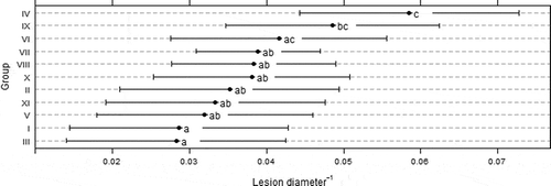
Fig. 4. Pathogenicity of Monilinia spp. isolate groups. Lesion growth was assessed after 4 days of incubation at 22 ° C with 12 h light regime. Different letters correspond to significant differences of means (P > 0.05) based on false discovery rate test.

Reprints and Corporate Permissions

Please note: Selecting permissions does not provide access to the full text of the article, please see our help page How do I view content?

To request a reprint or corporate permissions for this article, please click on the relevant link below:

Academic Permissions

Please note: Selecting permissions does not provide access to the full text of the article, please see our help page How do I view content?

Obtain permissions instantly via Rightslink by clicking on the button below:

If you are unable to obtain permissions via Rightslink, please complete and submit this Permissions form. For more information, please visit our Permissions help page.