
Figures & data
Table 1. Primer sets, sequences and amplicon size used to identify bacterial pathogens using single-colony PCR or multiplex PCR using crude plant extracts as the template.
Fig. 1 (Colour online) Symptoms of bacterial diseases observed during field surveys conducted in Alberta in 2012, with corresponding pathogens isolated from leaves in the lab. (a) Large yellow halos surrounding small necrotic lesions typical of halo blight; Pph isolated; (b) Brown spot symptoms showing spotty brown necrotic lesions with slight yellow margins; Pss isolated. (c) Symptoms typical of brown spot, but both Pph and Pss were isolated; (d) Early symptoms of common blight with brown necrotic area and yellow margins; both Xff and Pph isolated; (e) Foliar symptoms of bacterial wilt with spreading brown, senescent lesions and slight yellow margins; both Cff and Pph were isolated; and (f) symptoms typical of abiotic injury yielding fluorescent Pseudomonads only.
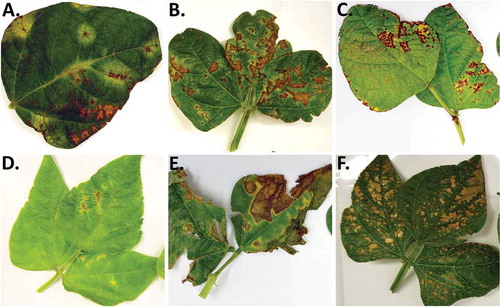
Table 2. Occurrence and severity of bacterial disease symptoms observed during surveys of commercial dry bean fields in southern Alberta.
Fig. 2 Change in bacterial blight incidence (a) and severity (b) over the growing season in 2012.
Table 3. Frequency of isolation (%) of bacterial pathogens from bean tissue samples collected in Alberta during 2012 and 2014 using three different identification methods: colony morphology on milk-tween agar, single colony PCR and PCR from plant extract.
Table 4. Identification of Xanthomonas-like bacteria isolated from bean leaf samples using 16S ribosomal sequences and length of lesions on bean stems ‘CDC Sol’ following injection of bacterial suspensions.
Table 5. Race characterization of Pseudomonas syringae pv. phaseolicola (Pph) isolates collected from commercial dry bean fields in western Canada in 2011–2014, as determined by reaction on the set of eight differential bean lines.
Table 6. Response of common blight resistant dry bean breeding lines from the AAFC-Morden bean breeding programme to P. syringae pv. phaseolicola isolate AB2012.002 (race 2) collected in Alberta, as determined in greenhouse trials.
Table 7. Halo blight disease reaction of selected bean genotypes from commonly grown commercial market classes to P. syringae pv. phaseolicola isolate AB2012.002 (race 2) collected in Alberta, as determined in greenhouse trials.