Figures & data
Fig. 1 (Colour online) Symptoms of field-infected okra pods and the isolated fungus. (a) Infected pod displaying white mycelium growth and sclerotial development. (b) Pure culture of the isolated fungus showing white fluffy mycelium and rings of sclerotia. (c) Hyaline mycelium of S. sclerotiorum. (d) Stalk of mature apothecium.
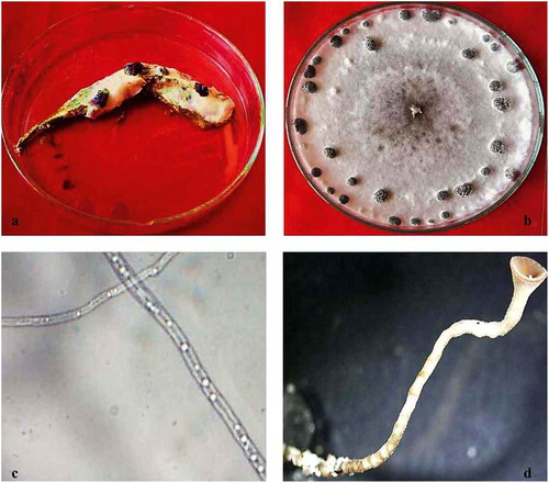
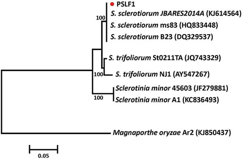
Fig. 2 (Colour online) Phylogenetic tree constructed with the ITS-5.8S rDNA sequence of the isolate from this study (PSLF1), and other species of Sclerotinia retrieved from GenBank. Magnaporthe oryzae was used as the out-group taxon. The bar indicates nucleotide substitutions per site. Numbers of bootstrap support values ≥ 50% based on 1000 replicates.