Figures & data
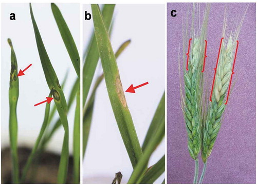
Fig. 1 Head and leaf blast symptoms on triticale plants under field conditions; (a) typical eye-shaped lesions on triticale leaf with white centre surrounded by brown margin; (b) & (c) partially bleached triticale spikes with dark grey to black pigmented infection points on rachis (inset)
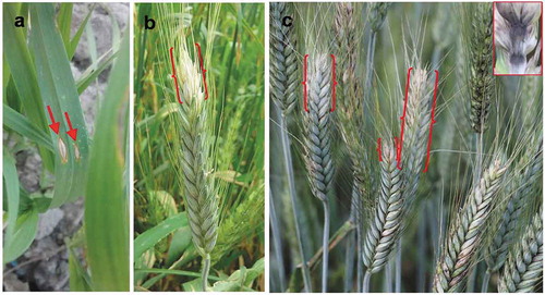
Fig. 2 Vegetative and reproductive growth of Magnaporthe oryzae (pathotype Triticum) under laboratory conditions; (a) ash coloured conidial growth of the fungus on the incubated rachis; (b) 2-septed hyaline pear-shaped conidia under compound microscope (magnification 400X); (c) single-spore colony of both isolates – i. TrBWMRI-19M6, ii. TrBWMRI-19M7 – on potato dextrose agar medium; and (d) sporulation of both isolates – i. TrBWMRI-19M6, ii. TrBWMRI-19M7 – on oatmeal agar medium
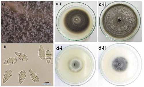
Fig. 3 PCR analysis of triticale blast isolates and some other blast isolates from different hosts with an ITS primer set (ITS5 and ITS4)
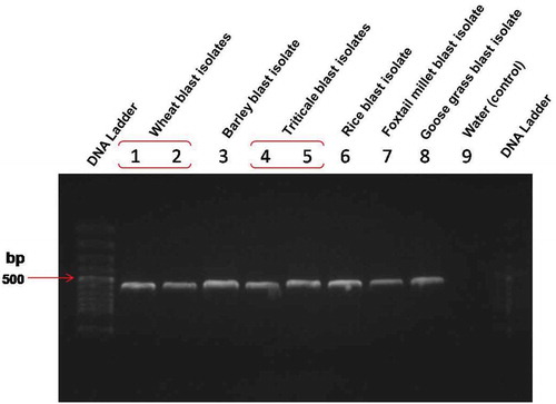
Fig. 4 PCR analysis of triticale blast isolates including some other hosts (wheat, barley, rice, foxtail millet, and goose grass) with MoT3 primers for confirming the Magnaporthe oryzae pathotype Triticum in a molecular detection assay
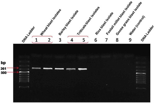
Fig. 5 Phylogenetic tree of Magnaporthe oryzae from triticale plants in Bangladesh and related species based on a neighbour joining likelihood analysis of a combined internal transcribed spacer (ITS) dataset. The ITS sequence data for the other nine isolates were retrieved from the NCBI GenBank database. Numbers beside each branch represent bootstrap values obtained after a bootstrap test with 1,000 replications. The bar indicates the number of nucleotide substitutions. The two triticale blast isolates are indicted by the red diamond shape. This analysis was performed with MEGA X software

Fig. 6 Pathogenicity confirmation of the M. oryzae pathotype Triticum fungus (isolate TrBWMRI-19M6) by fulfiling the Koch’s postulates; (a) round to oval-shaped water-soaked lesions developed on the seedling leaves 2–3 days after inoculation with Magnaporthe oryzae (TrBWMRI-19M6); (b) grey centred elongated lesion on seedling leaf at 7 days post-inoculation; and (c) partially bleached head blast symptoms developed 10–12 days after inoculation