Figures & data
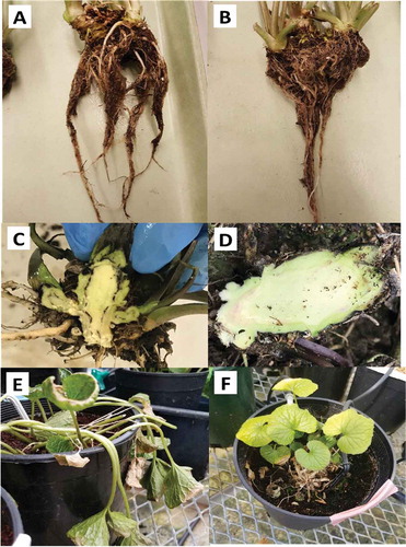
Fig. 1 (Colour online) Natural progression of disease symptoms on greenhouse grown wasabi plants infected by verticillium wilt. (a, b) Wilting of foliage under warm conditions in the greenhouse. (c, d, e) Blackening of the vascular tissue observed in diseased plants. (f) V-shaped necrotic lesions observed on some leaves. (g) Necrosis of leaf margins. Isolations made from symptomatic rhizomes and leaves yielded colonies of Verticillium.
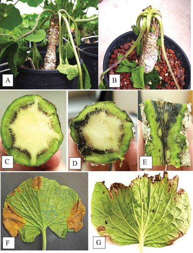
Fig. 2 (Colour online) Colonies of Verticillium isaacii grown on PDA. (a) Isolates form velvety, white colonies after 7 days. B) Colonies darken to black as time progresses to 14 days. (c) Some isolates, such as ECB4, produce yellow pigments in culture after 7 days. (D-F) Morphology of conidiophores and conidia of Verticillium isaacii. (d) Verticilliate conidiophores with 3 to 5 phialides. (e) Abundant hyaline conidia. (f) Dark microsclerotia about 1 mm in diameter. Scale bars = 25 µm
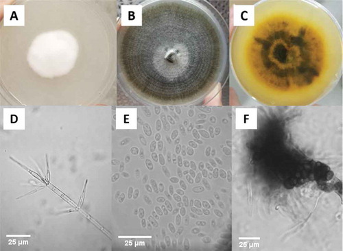
Fig. 3 Phylogenetic analysis of isolate NFIS4923 identified it as Verticillium isaacii/tricorpus based on analyses using ITS-ITS 2 primers. The evolutionary history was inferred using the Neighbor-Joining method. The percentage of replicate trees in which the associated taxa clustered together in the bootstrap test (1000 replicates) are shown next to the branches. The tree is drawn to scale, with branch lengths in the same units as those of the evolutionary distances used to infer the phylogenetic tree. The evolutionary distances were computed using the Maximum Composite Likelihood method and are in the units of the number of base substitutions per site. The analysis involved 31 and nucleotide sequences. All positions containing gaps and missing data were eliminated. There were a total of 448 positions in the final dataset. Evolutionary analyses were conducted in MEGA7 (Kumar et al. Citation2016)
Fig. 4 Phylogenetic analysis of isolate NFIS4923 identified it as Verticillium isaacii based on analyses using actin primers (Inderbitzin et al. Citation2013). The evolutionary history was inferred using the Neighbour-Joining method. The percentage of replicate trees in which the associated taxa clustered together in the bootstrap test (1000 replicates) are shown next to the branches. The tree is drawn to scale, with branch lengths in the same units as those of the evolutionary distances used to infer the phylogenetic tree. The evolutionary distances were computed using the Maximum Composite Likelihood method and are in the units of the number of base substitutions per site. The analysis involved 30 nucleotide sequences. All positions containing gaps and missing data were eliminated. There were a total of 317 positions in the final dataset. Evolutionary analyses were conducted in MEGA7 (Kumar et al. Citation2016)
Fig. 5 Verticillium isaacii grown for 7 days at temperatures from 5° to 35°C. Graph displays average diameter of colonies of two isolates based on two trials. Error bars = 95% CI
Fig. 6 (Colour online) Detached and wounded tissues inoculated with a mixed-isolate suspension of Verticillium isaacii resulting in symptom development. (a) Chlorotic lesions on leaves developed from the three points of inoculation compared to a wounded noninoculated leaf in (b). (c) Rhizome blackening from the point of inoculation compared to a healthy control (d). (e) Blackening of petiole tissue at the point of inoculation compared to a healthy petiole (f)
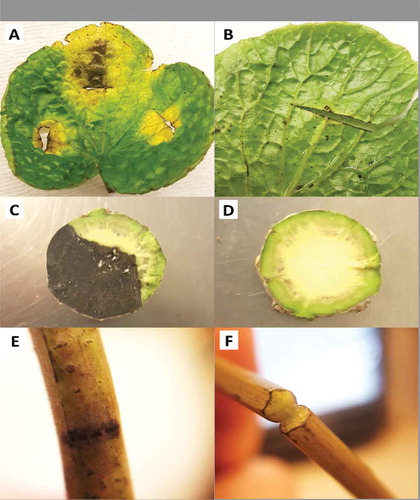
Fig. 7 (Colour online) Symptoms on plants inoculated with a mixed-isolate mycelial suspension of Verticillium isaacii (a) Inoculated rhizome shows visibly sparser roots compared to a control plant (b) after 3 months. (c) All inoculated plants had some degree of vascular blackening in the rhizome tissues while controls were asymptomatic (d). (e) Wilting occurred in one of the three inoculated plants while the controls remained unwilted (f)