Figures & data
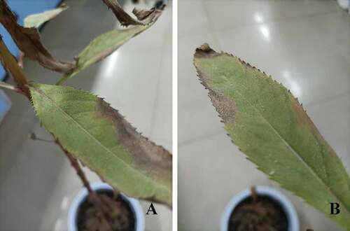
Fig. 1 Signs and symptoms of powdery mildew disease on Impatiens balsamina.
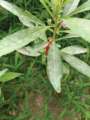
Table 1. Podosphaera sp. and their corresponding hosts
Fig. 2 Fungal structures of P. xanthii on host leaf surface. The structures were stained with trypan blue.
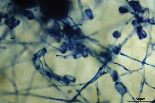
Fig. 3 PCR detection of the powdery mildew on I. balsamina samples collected in 2021 (A) and 2020 (B). M: marker; 1–4: diseased samples.
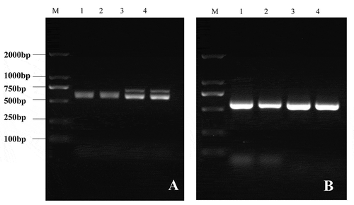
Fig. 4 Phylogenetical analysis of P. xanthii and Podosphaera sp. The neighbour joining tree was constructed in MEGA software with 1000 bootstrap replicates and p-distance method. The bar indicates a distance of 0.020. Red dot highlights P. xanthii. Arthrocladiella mougeotii and Erysiphe sp. were included as outgroup.
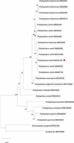
Fig. 5 P. xanthii infected I. balsamina and signs of powdery mildew 10-days post inoculation.