Figures & data
Figure 1. The working principle of the polymerase/endonuclease assisted colorimetric microRNA detection method.
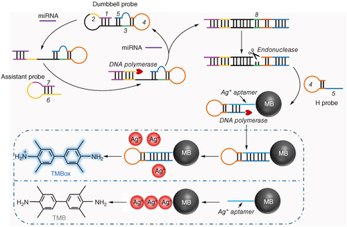
Figure 2. The feasibility of target recycling and colorimetric approach for miRNA detection. (A) SYBR Green I signals of the DP with different incubation time. (B) Fluorescence intensities of the dumbbell probe during the target recycling process. (C) Fluorescence intensities of the FAM labeled assistance probe during the target recycling process. 1, AP before assembly to hairpin structure; 2, AP; 3, AP + DP; 4, AP + DB + miRNA. (D) Fluorescence intensities of the FAM (carboxyfluorescein) labeled aptamer before and after construction of the aptamer@MB. (E) Absorbance of the approach when several experimental parameters existed or not.
AP: Assistance probe; DP: Dumbbell probe.
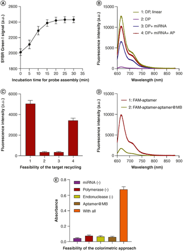
Figure 3. Optimization of experimental parameters. Absorbance of the approach when detecting miRNA with different incubation time (A), different enzyme concentrations (B), different amounts of the assistance probe (C) and the volume of the aptamer@MB complex (D).
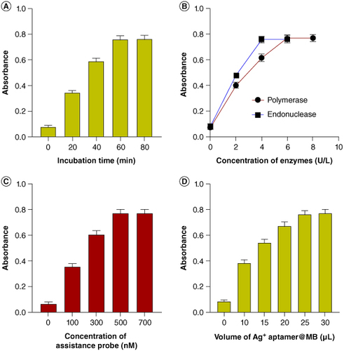
Figure 4. Sensitivity of the approach. (A) Absorbance of the approach when detecting different concentrations of target miRNA. (B) Correlation between the absorbance anf the concentration of target miRNA.
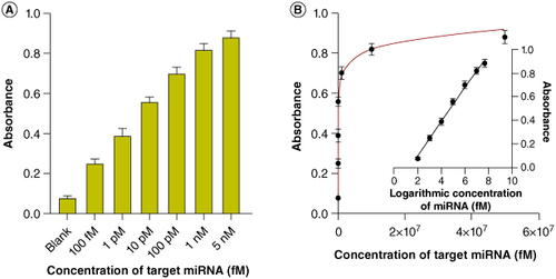
Figure 5. Selectivity of the approach. (A) Absorbance of the approach when detecting different miRNAs. (B) Absorbance of the approach and traditional method when detecting mismatched sequences.
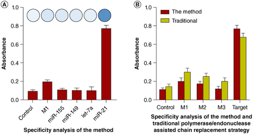
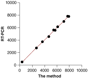
Figure 6. Biological sample application of the approach.