
Figures & data
Table 1. Subjects’ characteristics.
Figure 1. The time schedule for each experiment.
The bars are colored according to the time of day: white, daytime; light gray, nighttime and dark gray, sleep time. The slim, horizontal shaded bars indicate the measurement times for resting energy expenditure (REE) and autonomic nervous activity (ANA) around mealtimes.
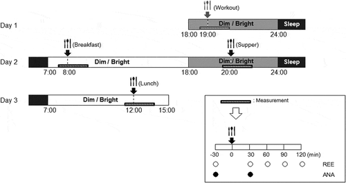
Figure 2. Changes in diet-induced thermogenesis (DIT) following meal intake under three light conditions.
Changes in DIT with (a) the DD treatment (dim light exposure throughout the experiment); (b) the BD treatment (bright light exposure during the daytime and dim light exposure during the nighttime) and (c) the BB treatment (bright light exposure throughout the experiment except for sleep time). †: p < 0.1.
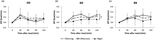
Table 2. Contents and nutrient composition of the meal.
Table 3. Diet-induced thermogenesis (DIT) and autonomic nerve activities (ANA) at different times of the day under different light conditions.