Figures & data
Table 1. Clinical characteristics.
Table 2. Associations with the scaling exponent α.
Figure 1. Average detrended fluctuation functions F(n) from locomotor activity data of 72 healthy controls, 68 NFMA patients and 22 craniopharyngioma patients. Data are shown on log-log plots and curves are vertically shifted in [A] for better visualization of differences between groups (vertical offset does not alter the slope). The scaling behavior can be separated in three time scale regions based on two breakpoints at ~1.5 h and ~10 h. The break points can be seen more clearly when F(n) is divided by time scale n in [B], indicating the change in F(n) per time unit n. The straight lines represent perfect scale-invariance α = 1.00 [A] or α’ = 0.00 [B].
![Figure 1. Average detrended fluctuation functions F(n) from locomotor activity data of 72 healthy controls, 68 NFMA patients and 22 craniopharyngioma patients. Data are shown on log-log plots and curves are vertically shifted in [A] for better visualization of differences between groups (vertical offset does not alter the slope). The scaling behavior can be separated in three time scale regions based on two breakpoints at ~1.5 h and ~10 h. The break points can be seen more clearly when F(n) is divided by time scale n in [B], indicating the change in F(n) per time unit n. The straight lines represent perfect scale-invariance α = 1.00 [A] or α’ = 0.00 [B].](/cms/asset/d00ff538-39bb-41a6-9b12-74008a6d6b50/icbi_a_1407779_f0001_b.gif)
Figure 2. Scaling exponent α in 72 healthy controls, 68 NFMA patients and 22 craniopharyngioma patients (CP), obtained from detrended fluctuation analysis, separated in three time scale (n) regions of the 24-h analysis period. The dotted lines represent too much randomness or white noise at α = 0.5, too much regularity or red noise at α = 1.5, and the delicate balance between the two in healthy systems known as complex scale invariance or pink noise at α = 1.0. Bars represent mean ± SD.
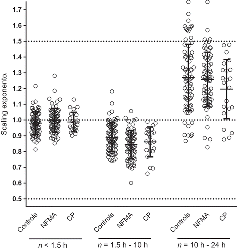
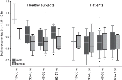
Figure 3. Scaling exponent α2 (times scales 1.5–10 h) in 72 healthy subjects and 90 patients (NFMA and craniopharyngioma). Data are separated by gender and in four age groups. In healthy subjects, group size for each age group in men is 3, 8, 11 and 16, and for women 3, 7, 19 and 5. For patients, group size for each age group in men is 3, 9, 25 and 13, and for women 4, 6, 21 and 9. The dotted line represents complex scale invariance with α = 1.0. Boxes display the 25th, 50th and 75th percentile, bars the 10th and 90th percentile and dots the outliers.