
Figures & data
Figure 1. Dermatitis herpetiformis. (A) Blisters on the elbows and knees. (B) Fresh, small blisters on the elbow. (C) Excoriated blisters and scars on the elbow, a typical clinical picture caused by scratching the lesions.
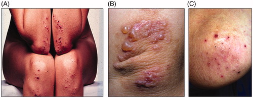
Figure 2. Immunofluorescence findings in dermatitis herpetiformis. Pathognomonic granular IgA deposits in the basal membrane zone between epidermis and dermis, and also deeper in the dermal tissue.
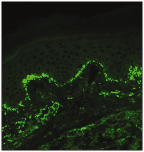
Table 1. Comparison between dermatitis herpetiformis and coeliac disease.