Figures & data
Figure 1. Illustration of the used computed tomopraghy angiography (CTA) classification of the internal carotid artery (ICA) stenosis and an example of an ulcerative CP. Images A–D represent CTA axial plane source images showing the different classifications of the ICA stenosis. Images E – H represent the corresponding oblique sagittal maximum intensity projection reformat images. All the images visualize a right sided ICA stenosis. A and E = bulky calcification; B and F = soft plaque (delineated); C and G = ulcerative plaque (arrow marks the pouch of a plaque ulceration); D and H = calcified nodules.
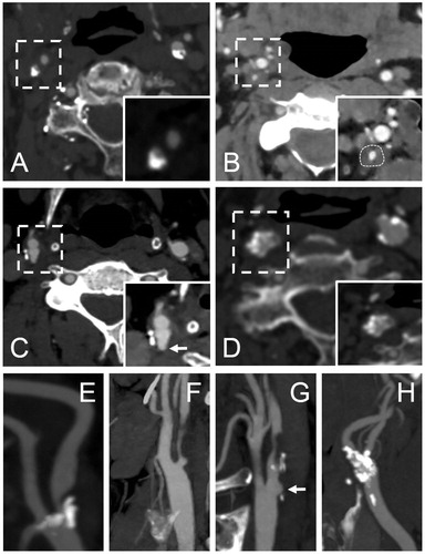
Figure 2. Morphologic characteristics and their evaluation criteria. Several characteristics were often observed in one specimen.
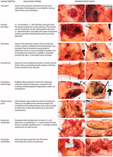
Figure 3. HeCES2 study patients and carotid plaques – origin of symptoms. 413 (83%) of the 500 study patients had experienced a cerebrovascular symptom, 67 (13%) were asymptomatic and 20 (4%) had an unspecified symptom, that is not possible to determine whether it represented a cerebrovascular or an alternative symptomatology such as epileptic, migrainous or musculoskeletal. Among the 413 study patients with a cerebrovascular symptom, the operated CP was determined to be symptom-causing in 324 (79%) patients while in 35 (8%) patients it was deemed that the CP was innocent and the cerebrovascular symptom represented another aetiology or vascular territory. In another 54 (13%) patients, coinciding competing aetiologies of cerebrovascular symptoms prevented to declare whether the CP was symptom-causing or not. Among the symptom-causing CPs, 161 (50%) had caused a stroke and 163 (50%) a TIA, and further 137 (85%) of the strokes were hemispheric and 24 (15%) retinal artery occlusions, whereas 79 (48%) of the TIAs were hemispheric and 84 (52%) were amaurosis fugax.
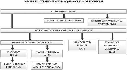
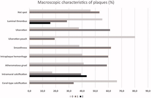
Figure 4. The proportion of macroscopic characteristics in carotid plaques. All characteristics were divided into two categories: 0 = not present or 1 = present, except intramural calcification and luminal thrombus, that had three different categories: 0 = not present, 1 = moderately present, 2 = abundantly present.