
Figures & data
Table 1. Baseline demographic and clinical characteristics of participants by sex.
Figure 1. Estimated average and standard deviation of cf-PWV, ba-PWV and CAVI in subjects with and without vascular and renal TOD, and LVH. p-value: differences among groups. TOD: target organ damage; LVH: left ventricular hypertrophy; cf-PWV: carotid-femoral pulse wave velocity; ba-PWV: brachial-ankle pulse wave velocity; CAVI: cardio-ankle vascular index.
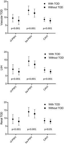
Figure 2. Estimated average and standard deviation of cf-PWV, ba-PWV and CAVI in subjects with and without vascular and renal TOD, and LVH. Adjusted for age (years) and sex (0= female, 1 = male). p-value: differences among groups. TOD: target organ damage; LVH: left ventricular hypertrophy; cf-PWV: carotid-femoral pulse wave velocity; ba-PWV: brachial-ankle pulse wave velocity; CAVI: cardio-ankle vascular index.
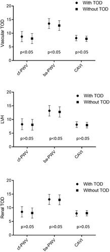
Table 2. Bivariate correlations of structure and function parameters with cardiovascular risk factors.
Table 3. Regression analysis of vascular structure and function parameters with vascular, cardiac, and renal parameters.
Figure 3. A. Logistic regression analysis adjusted by age (years), smoking (years), sex, alcohol (gr), total physical activity (hour/week) and Mediterranean Diet (points), OR of cf-PWV, ba-PWV and CAVI with vascular and renal TOD, and LVH. B. Logistic regression analysis adjusted by age (years), smoking (years), sex, alcohol (gr), total physical activity (hour/week), Mediterranean Diet (points), SBP (mmHg), BMI (kg/m2), FPG (mg/dl) and triglycerides (mg/dl), OR of cf-PWV, ba-PWV and CAVI with vascular and renal TOD, and LVH. TOD: target organ damage; LVH: left ventricular hypertrophy;cf-PWV: carotid-femoral pulse wave velocity; ba-PWV: brachial-ankle pulse wave velocity; CAVI: cardio-ankle vascular index.
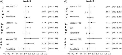
Figure 4. Receiver operating curve of cf-PWV, ba-PWV and CAVI to identify vascular TOD (a), LVH (b) and renal TOD (c). Areas under the ROC curves are summarised in . TOD: target organ damage; LVH: left ventricular hypertrophy; cf-PWV: carotid-femoral pulse wave velocity; ba-PWV: brachial-ankle pulse wave velocity; CAVI: cardio-ankle vascular index.
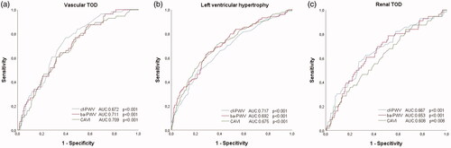
Table 4. Area under receiver operating characteristics curve and cut-off points sensitivity and specificity for the vascular and renal TOD, and LVH.