
Figures & data
Figure 1. Expression of miR-492 in NSCLC tissues and cell lines. (A) The expression of miR-492 was increased in NSCLC tissues. (B) The expression level of miR-492 was increased in four NSCLC cell lines compare with normal NHBE cell lines. (C) NSCLC patients with high miR-492 expression had worse survival compared with low miR-492 expression, illustrating that NSCLC patients with high miR-492expression were associated with worse prognostic survival (Log-rank p = .0007) (***p < .001).
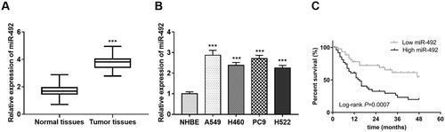
Table 1. Clinicopathological characteristics in patients with NSCLC.
Table 2. Multivariate Cox regression analysis to adjust the association of miR-492 with NSCLC patients’ survival prognosis.
Figure 2. miR-492 inhibitor suppresses cell proliferation, migration and invasion of NSCLC cells. (A) After transfection with miR-492 inhibitors, miR-492 expression levels in A549 and PC9 cell lines decreased compared with inhibitor NC (all p < .001). (B, C) Silencing of miR-492 suppressed cell proliferation in A549 and PC9 cell lines compared with inhibitor NC, and the difference gradually became significant with time (48 h: all p < .01; 72 h: all p < .001). (D, E) Silencing of miR-492 significantly inhibited cell migration and invasion in A549 and PC9 cell lines compared with inhibitor NC. Transwell images observed by inverted microscopy showed that the number of cell migration and invasion was the least in A549 and pC9 cells transfected with miR-492 inhibitor (all p < .001) (**p < .01; ***p < .001).

Figure 3. UTMD enhances cell transfection efficiency of miR-492. (A, B) UTMD could significantly enhance the inhibition of miR-492 inhibitor on the expression level of miR-4284 in A549 and PC9 cell lines, and the efficiency of transfection increased (***p < .001; #p < .05; ##p < 0.01; *compare with control, ##compare with miR-492 inhibitor).
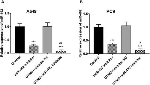
Figure 4. Effects of UTMD-mediated miR-492 inhibitor transfection on NSCLC cell proliferation, migration and invasion. (A) UTMD-mediated miR-492 inhibitor further inhibited cell proliferation compared with transfection of miR-492 inhibitor alone. (B, C) UTMD-mediated miR-492 inhibitor further inhibited cell migration and invasion compared with a single miR-492 inhibitor (*p < .05; **p < .01; ***p < .001; #p < .05; *compare with control, #compare with miR-492 inhibitor).
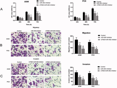
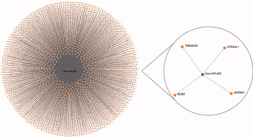
Figure 5. Target genes of miR-492 predicted by the miRWalk database.
Data availability statement
The data used to support the findings of this study are available from the corresponding author upon reasonable request.